Axial T2WI at the level of suprasellar cistern.
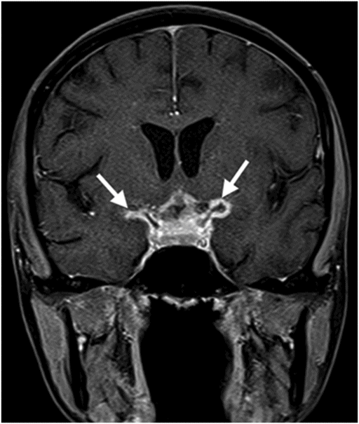
Coronal enhanced T1WI at the level of the cavernous sinuses.
Tubercular Vasculitis
Primary Diagnosis
Tubercular vasculitis
Differential Diagnoses
HIV cerebrovascular disease
Neurosyphilis (meningovascular disease)
Imaging Findings
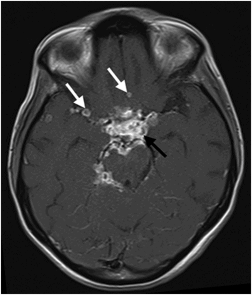
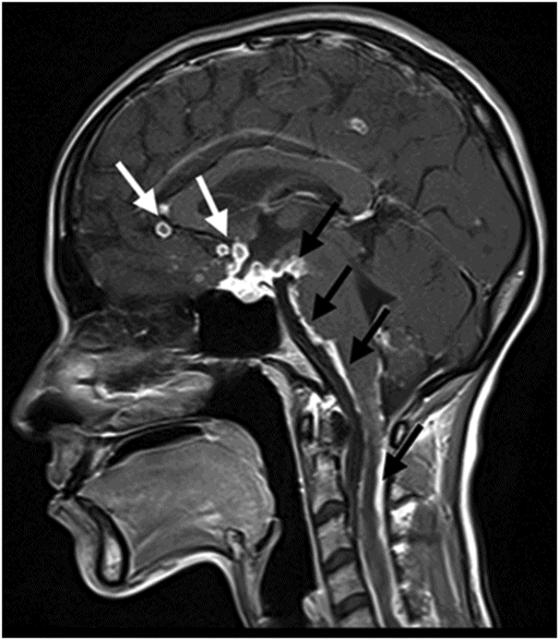
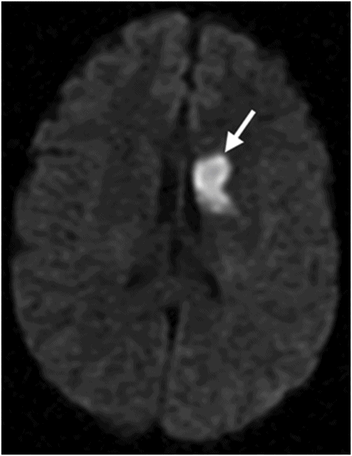
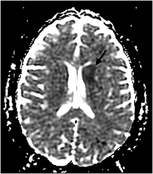
Fig. 45.1: Axial T2WI at the level of suprasellar cistern showed hypointense material at the suprasellar cistern surrounding the optic chiasm and tracts (black arrow). At this level there were small hyperintense foci in the midbrain (white arrows), in keeping with inflammatory changes. Fig. 45.2: Axial enhanced T1WI at the level of the basal cisterns, showed enhancing material in the suprasellar, interpeduncular, and perimesencephalic cisterns, in keeping with exudative material due to basilar meningitis (black arrow). Also, note the small, rounded ring-enhancing lesions in the basifrontal region and in the right sylvian fissure, in keeping with granulomas (white arrows). Fig. 45.3: Sagittal enhanced T1WI at the level of the midline showed the same enhancing material in the sellar, suprasellar, prepontine, and perimedullary cisterns. Note that this material also diffusely coated the surface of the spinal cord, in keeping with extensive granulomatous meningitis (black arrows). Please note the multiple, small, rounded ring-enhancing lesions in the frontal region and in the suprasellar cistern, in keeping with granulomas (white arrows). Fig. 45.4: Coronal enhanced T1WI at the level of the cavernous sinuses showed enhancing material in the sellar, suprasellar cisterns, and surrounding the proximal portions of the middle cerebral arteries, and supraclinoid internal carotids, reflecting perivascular inflammatory changes (arrows). Fig. 45.5: Axial DWI and Fig. 45.6: ADC map at the level of the lateral ventricles showed a focus of true restricted diffusion at the left caudate nucleus, in keeping with acute ischemic changes.
Discussion
The findings of extensive exudative enhancing material in the basal cisterns and posterior fossa cisterns, which coats the cervical spinal cord ( in keeping with basilar meningitis), in association with ring-enhancing lesions in the subarachnoid space at those same levels (granulomas), perivascular inflammatory changes, and acute ischemic changes in a patient with upper lung lesions, which are positive acid-fast bacilli on Ziehl-Neelsen stain, are highly suspicious for tuberculous meningitis (TBM) associated with vasculitis complicated with parenchymal infarcts.
Human immunodeficiency virus infection is a worldwide epidemic disease with commonly reported neurologic complications. Despite its universal distribution, the presence of HIV-related cerebrovascular disease (CVD) is an uncommon event. Forty to fifty percent of AIDS patients can have a neurologic complication, but less than 2% have a stroke syndrome, more commonly ischemic infarcts than intracerebral hemorrhages. Cerebral infarcts are usually associated with non-bacterial thrombotic endocarditis or concomitant opportunistic CNS infection, and intracerebral hemorrhages are usually associated with thrombocytopenia, primary CNS lymphoma, and metastatic Kaposi sarcoma. Owing to the absence of HIV infection, and the fact that the typical imaging findings of HIV/AIDS-related ischemic changes are not associated with basilar meningitis and intracranial granulomas, this entity is less likely in this patient.
Syphilis is a sexually transmitted infectious and disabling disease caused by the spirochete Treponema pallidum, with increased incidence due to the HIV/AIDS epidemic. Neurologic complications of syphilis or neurosyphilis (NS) are well known and can be quite severe. Neurosyphilis occurs in about 10% of untreated syphilitics and typically manifests during the secondary or later stages of the disease. It can compromise all components of the nervous system, including the spinal cord, as well as cranial and peripheral nerves, meninges, and CNS blood vessels.
Meningovascular syphilis (MVS) is a distinct form of NS characterized by a meningo-encephalopathic syndrome with superimposed cerebrovascular or myovascular events. Meningovascular syphilis occur in 10% of NS patients and in 3% of syphilis cases. Meningovascular syphilis involves an infection-associated inflammatory arteriopathy resulting in injury to the blood vessels of the leptomeninges, brain, and spinal cord, leading to infarctions. Meningovascular syphilis-related cerebral infarcts are non-specific and dependent upon the arterial territory involved. Acute spinal cord infarcts resulting from MVS may clinically present in various forms of myelopathy, including hemiparaplegic syndrome (Brown-Séquard hemiplegia) or transverse myelitis. The absence of a history of HIV and syphilis, and the lack of association with granulomas and basilar meningitis, makes the possibility of MVS in this particular patient a less likely diagnosis.
Tuberculosis (TB) is an infectious disease caused by Mycobacterium tuberculosis bacilli. Tuberculosis is still prevalent in many regions of the world and the incidence of TB has markedly increased in recent years, particularly in conjunction with AIDS and the emergence of drug-resistant strains of the bacillus. The lungs are the most commonly affected site, but many organs and tissues may be targeted.
Tuberculous involvement of the CNS is an important and serious type of extrapulmonary involvement, occurring in around 10% of TB patients. Central nervous system TB can occur in 19% of the patients with coexisting TB and HIV.
It is believed that the bacilli reach the CNS hematogenously, secondary to disease elsewhere in the body. Tuberculosis develops in the CNS in two stages. Initially small tuberculous lesions (Rich’s foci) develop in the CNS, either during the stage of bacteremia of the primary TB infection or shortly afterwards. These initial granulomatous inflammatory reactions may occur in the meninges, subpial, or subependymal surface of the brain or the bone covering of the brain or the spinal cord, and may remain dormant for years after initial infection.
Later, rupture and growth of these small tuberculous lesions produces development of various types of CNS TB including parenchymal and leptomeningeal tuberculomas, abscesses, cerebritis, vasculitis, infarction, meningitis, and osteomyelitis. The specific stimulus for rupture or growth of Rich’s foci is not known, although immunologic mechanisms are believed to play an important role.
The most common forms of intracranial TB are: meningeal (leptomeningeal and pachymeningeal) and parenchymal (tuberculomas, tuberculous abscess, tuberculous cerebritis, and tuberculous encephalopathy). Complications of TBM are tuberculous vasculitis, hydrocephalus, and cranial nerve involvement.
Intracranial vasculitis is a common finding in patients dying from TBM and a major factor contributing towards residual neurologic deficits. In patients with infarcts, TBM is reportedly up to three times more fatal than in those patients without infarcts. In survivors, the extent of ischemic cerebral damage is an important determinant of disability.
The pathophysiology remains unclear, but it is believed that the basal exudate of TBM causes inflammatory changes in the vessels that predominantly involve the circle of Willis. At first, the vessel wall is involved, and later the lumen, leading to complete occlusion by reactive subendothelial cellular proliferation constituting panarteritis tuberculosa. Middle cerebral and lenticulostriate arteries are the most common vessels involved.
Stroke is a common complication of tuberculous vasculitis in the context of TBM. Most infarcts in TBM are because of hemodynamic hypoperfusion due to a variable combination of vasospasm, intimal proliferation, and thrombosis of cerebral blood vessel walls. It is regarded as a poor prognostic predictor of TBM. The majority of the strokes in TBM are usually small, multiple, bilateral, and located in the basal ganglia, especially in the tubercular zone, which comprises the caudate, anterior thalamus, anterior limb, and genu of the internal capsule, corresponding to the deep sylvian region. Cortical stroke can also occur because of the involvement of the proximal portion of the middle, anterior, and posterior cerebral arteries, as well as the supraclinoid portion of the internal carotid and basilar arteries.
The conventional angiographic features include narrowing of arteries at the base of the brain, and narrowed or occluded small or medium-sized arteries with early draining veins. Leptomeningeal vessels over the convexities of the brain may be stretched because of internal hydrocephalus or brain swelling. Computed tomography or MR angiogram reveal small segmental narrowing, uniform narrowing of large segments, irregular beaded appearance of vessels, or complete occlusion. Although the cerebral vasculature can be visualized by MRA, direct angiography remains the gold standard for imaging the vascular lumen.
Magnetic resonance imaging is more sensitive and detects a greater number of infarcts and hemorrhagic transformation of infarcts than CT does. On MR imaging, ischemic lesions are typically seen as hyperintensities on T2-weighted images. Diffusion-weighted imaging helps in early detection of ischemic lesions and in delineating the extent of infarction, which is of value in the management and outcome of patients.
Tuberculous vasculitis is an important cause of morbidity and mortality in patients with TBM. Angiographic and MR imaging plays a crucial role in diagnosis because of its inherent sensitivity and specificity in detecting vessels narrowing and CNS strokes. The knowledge of this complication may help in better management of patients with CNS TB.
Stay updated, free articles. Join our Telegram channel

Full access? Get Clinical Tree


