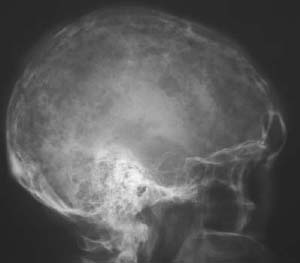
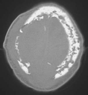
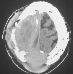
CASE 47 George Nomikos, Anthony G. Ryan, Peter L. Munk, and Mark Murphey A 49-year-old man was admitted to the hospital who was unconscious and had a fever. Figure 47A Figure 47B Figure 47C A lateral radiograph (Fig. 47A) shows diffuse osseous destruction throughout the skull in a moth-eaten/permeative pattern. The axial unenhanced CT image (Fig. 47B) also demonstrates this diffuse osseous infiltration and destruction throughout the skull. Several larger lytic areas with associated soft-tissue masses are also identified. No mineralized matrix is seen. The contrast-enhanced CT (Fig. 47C) obtained at a higher level shows two enhancing soft-tissue masses arising from the skull and causing compression of the underlying brain parenchyma. Multiple myeloma. Myeloma, a lymphoproliferative malignancy of monoclonal plasma cells, is the most common primary malignancy of bone, with an incidence of ~50 cases/100,000 people per year who have reached an age of 80 years. The majority of patients (55 to 60%) have multiple myeloma, which is characterized by multiple sites of involvement. The solitary form of the disease (solitary plasmacytoma) accounts for ~25% of patients, although these patients usually go on to develop multiple lesions within 3 to 5 years. Because areas of hematopoietic marrow are predominantly affected, lesions primarily affect the axial skeleton. Although lesions are most commonly lytic, a sclerotic form of myeloma does exist. This sclerotic form of myeloma (Fig. 47D) is often, although not invariably, associated with POEMS syndrome: polyneuropathy, organomegaly, endocrinopathy, monoclonal gammopathy (usually immunoglobulin A or G [IgA or IgG]), and skin changes.
Multiple Myeloma
Clinical Presentation
Radiologic Findings
Diagnosis
Differential Diagnosis
Discussion
Background
Clinical Findings
Related posts:
Stay updated, free articles. Join our Telegram channel

Full access? Get Clinical Tree


