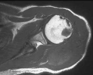
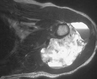
CASE 48 George Nomikos, Anthony G. Ryan, Peter L. Munk, Mark Murphey A 23-year-old man presented with intermittent aching in the left shoulder following a sports injury 1½ years prior to his visit to the hospital. Figure 48A Figure 48B Figure 48C The axial T1-weighted image (Fig. 48A) shows a soft-tissue mass around the proximal humerus. The mass is isointense to muscle. Note the focal area of marrow replacement in the humeral head compatible with osseous invasion. The mass is heterogeneously high signal intensity on the axial T2-weighted image (Fig. 48B) with no significant surrounding edema. Also note the fluid levels in the mass resulting from internal hemorrhage. The postcontrast T1-weighted image (Fig. 48C) demonstrates diffuse enhancement of the solid portion of the mass and peripheral enhancement of the cystic/hemorrhagic portion. The focal area of osseous invasion is also well illustrated on this coronal image. Synovial sarcoma.
Synovial Sarcoma
Clinical Presentation

Radiographic Findings
Diagnosis
Differential Diagnosis
Discussion
Background
Related posts:
Stay updated, free articles. Join our Telegram channel

Full access? Get Clinical Tree


