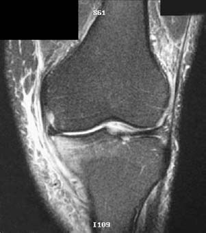
CASE 5 Anthony G. Ryan and Peter L. Munk A 26-year-old man presented with focal medial joint line pain, having received a heavy tackle to the outside of the leg while playing ice hockey. Figure 5A A coronal T2-weighted image (Fig. 5A) shows diffuse high signal and splitting of the medial collateral ligament (MCL). The intact proximal portion of the ligament is clearly seen. The distal half is poorly seen, attenuated, and discontinuous, and high signal fluid is shown internal and external to the ligament. MCL tear (grade III). None. Anatomically, the MCL consists of superficial and deep parts. The deep portion is inseparable from the true capsule of the joint, which in turn is adherent to the medial meniscus via meniscofemoral and meniscotibial ligaments. The superficial part of the MCL may be further divided into vertical and posterior oblique components. The vertical portion arises from the medial femoral epicondyle 5 cm above the joint line and inserts distally onto the medial tibial metaphysis 7 cm below the joint. The posterior oblique component extends from the femoral epicondyle to the posteromedial tibia and adjacent semimem-branosus tendon. Functionally, the ligament resists valgus forces at the knee and prevents excessive external rotation and anterior tibial translation. The vertical component remains constant in length throughout flexion, whereas the posterior oblique component becomes lax in flexion. Isolated MCL injury typically occurs with pure valgus stress without a rotatory component such as occurs in ice hockey injuries. MCL disruption is said to occur in up to 60% of skiing injuries, usually occurring when valgus stress is applied to the flexed knee. The MCL is the most frequent site of ligamentous injury (38%) found in association with closed fractures of the distal femoral shaft. The MCL is strong and when injured is frequently associated with internal derangements of the knee, particularly anterior cruciate ligament (ACL) and meniscal tears. When rotation is combined with valgus, the posterior oblique portion tends to be injured first, followed by the ACL, and then the remainder of the MCL. Patients frequently present with pain, swelling, and tenderness directly over the ligament. If an effusion develops immediately after the inciting injury, there is a high probability of an associated internal derangement, as the MCL is extra-articular. As a corollary, isolated tears should not produce an effusion. MCL integrity is assessed by valgus strain on the knee; however, the associated tenderness and muscle spasm limit the sensitivity of the examination, which is thus associated with a high false-negative rate. Valgus strain is applied at different angles of flexion: laxity at 30% implies isolated disruption of the MCL, whereas laxity in extension signifies a more extensive injury, as, for example, to the cruciate ligaments. O’Donoghue’s “unhappy” triad, consisting of an ACL tear, an MCL tear, and a medial meniscal tear with an associated lateral compartment bone bruise, occurs secondary to valgus with rotation. MCL injuries are graded on a three-point scale: Grade 1
Collateral Ligament Tear
Clinical Presentation

Radiologic Findings
Diagnosis
Differential Diagnosis
Discussion
Background
Etiology
Pathophysiology
Clinical Findings
Stages of Disease
![]()
Stay updated, free articles. Join our Telegram channel

Full access? Get Clinical Tree


