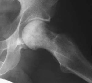
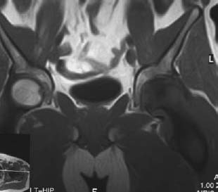
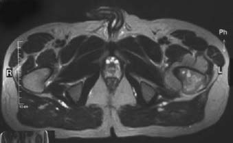
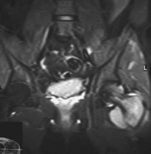
CASE 50 George Nomikos, Anthony G. Ryan, Peter L. Munk, and Mark Murphey A 38-year-old man presented with left hip pain. Figure 50A Figure 50B Figure 50C Figure 50D The frog-leg view of the left hip (Fig. 50A) shows sclerosis with some intermixed areas of lucency within the left femoral head and neck. The T1-weighted image (Fig. 50B) from the MRI examination shows marrow replacement in the femoral head and neck, as well as a large soft-tissue mass. On the T2-weighted image (Fig. 50C), marrow replacement is again seen in the proximal femur. The lesion is intermediate in signal intensity on the T2-weighted image. Also note the lack of obvious cortical destruction, despite the presence of a large soft-tissue mass. The large soft-tissue component with relative preservation of the cortex is also demonstrated on the STIR image (Fig. 50D). Primary lymphoma of bone.
Primary Lymphoma of Bone
Clinical Presentation
Radiologic Findings
Diagnosis
Differential Diagnosis
Discussion
Background
Related posts:
Stay updated, free articles. Join our Telegram channel

Full access? Get Clinical Tree


