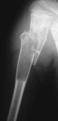
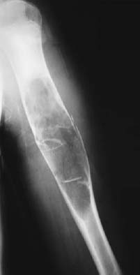
CASE 55 George Nomikos, Anthony G. Ryan, Peter L. Munk, and Mark Murphey A 12-year-old boy presented with a complaint of sudden onset of arm pain while playing. A radiograph was taken of the area (Fig. 55A). The next image (Fig. 55B) was obtained 3 years later. Figure 55A Figure 55B A single view of the left humerus (Fig. 55A) shows a pathologic fracture through a lytic lesion in the central medullary canal of the proximal humeral diaphysis. The lesion is mildly expansile and has a narrow zone of transition with mild surrounding sclerosis. There is no mineralized matrix in the lesion. The second image obtained 3 years later (Fig. 55B) shows a new pathologic fracture through the lesion. Both the humerus and the lesion had grown since the prior study. The lesion appears slightly more expansile on the current study than previously. Note the fragment of bone that has been displaced from the fracture site and has fallen into a dependent location of the lesion (fallen fragment). Simple (unicameral) bone cyst (SBC) with pathologic fracture. SBCs are nonneoplastic and represent the only true cystic lesion of bone. These lesions are benign and have an extremely low incidence of malignant transformation (there are rare reports of malignant transformation in the wall to osteosarcoma). SBCs are purely lytic lesions consisting of a fluid-filled cavity lined by a single layer of mesothelial-like cells. The etiology of these lesions is unknown, but various theories have been postulated, including (1) a defect of enchondral bone formation, (2) increased venous pressure, and (3) biochemical factors.
Unicameral Bone Cyst
Clinical Presentation


Radiologic Findings
Diagnosis
Differential Diagnosis
Discussion
Background
Clinical Findings
Stay updated, free articles. Join our Telegram channel

Full access? Get Clinical Tree


