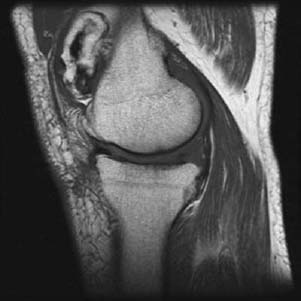
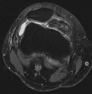
CASE 6 Anthony G. Ryan and Peter L. Munk Our patient presented with recurrent pain and locking after a remote trauma. Figure 6A Figure 6B A sagittal T1-weighted image (Fig. 6A) and an axial proton density (PD) fat-saturated image (Fig. 6B) show a very large irregular intra-articular loose body in the superomedial patellar recess secondary to degenerative osteoarthritis. Speckled low density scattered throughout the lesion is evident, consistent with calcifications. Chondromalacia patella is evident on the axial image. Articular surface irregularity and subchondral sclerosis are evident on the sagittal image. Intra-articular loose body. Solitary loose body: Multiple loose bodies: Emphasis will be placed on those conditions causing multiple small loose bodies, as the other conditions are dealt with at length elsewhere in the atlas. The etiology of large, solitary loose bodies is self-evident from the differential above. The so-called rice bodies associated with juvenile rheumatoid arthritis are composed of fragments of sloughed infarcted hypertrophied synovium, which are not typically calcified.
Intra-articular Loose Body
Clinical Presentation
Radiologic Findings
Diagnosis
Differential Diagnosis
Discussion
Background
Etiology
Related posts:
Stay updated, free articles. Join our Telegram channel

Full access? Get Clinical Tree


