PART IV Infection
CASE 67
Acute Pyogenic Osteomyelitis
Sam Y. Chun, Ali Islam, Alison Spouge, Anthony G. Ryan, and Peter L. Munk
Clinical Presentation
A 10-year-old girl presented with a 3-week history of thigh pain. Her blood work showed a mildly elevated erythrocyte sedimentation rate and normal white cell count.
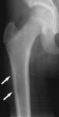
Figure 67A
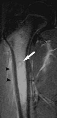
Figure 67B
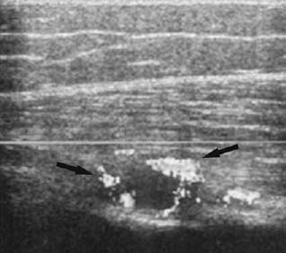
Figure 67C
Radiologic Findings
An anteroposterior radiograph of the femur (Fig. 67A) shows a subtle periosteal reaction (white arrows) along the lateral aspect of the diaphysis. Coronal fast inversion recovery (Fig. 67B) shows extensive high signal bone marrow edema in the proximal femoral metadiaphysis with a small spherical central ringlike low signal intensity consistent with very early abscess formation (arrow). A trace of fluid is also present in the periosseous soft tissues (arrowheads). Longitudinal sonogram (Fig. 67C) shows a small fluid collection (arrows) in the deep soft tissues adjacent to the proximal femur with a rim of hyperemia seen on color Doppler. The fluid collection was aspirated using ultrasound guidance, and subsequent cultures grew Staphylococcus aureus.
Diagnosis
Acute osteomyelitis.
Differential Diagnosis
- Eosinophilic granuloma
- Ewing’s sarcoma
- Lymphoma or leukemia
Discussion
Background
Osteomyelitis is an infection of bone. The term acute osteomyelitis is reserved for recent onset of osseous infection characterized by a relatively abrupt clinical presentation. Relapse of a previously documented infection or persistence of symptoms for more than 6 weeks is generally classified as subacute or chronic. Osteomyelitis occurs via three routes of contamination: hematogenous, spread from a contiguous source of infection, or direct implantation. In many instances the source of contamination cannot be identified, and occasionally more than one route may contribute to the infection. In children and adult populations with osteomyelitis, males appear to be more frequently affected than females; however, in infants, both sexes are affected with equal frequency.
Although hematogenous osteomyelitis can affect patients of all ages, it is considered a disease of childhood (3 to 15 years). The primary site of infection in children is the metaphysis of the long bones, particularly those bones involved in rapid growth, such as around the knee, the wrist, and the proximal humerus. In adults, the pelvis, spine, and small bones are more commonly affected. The primary focus of infection is sometimes evident (ear, skin, throat), but more often than not, no source can be found. Hematogenous osteomyelitis can involve single or multiple bones; in infants, multiple sites of infection are often seen due to septicemia from infected umbilical catheters.
In adults, osteomyelitis of the appendicular skeleton is more commonly due to spread from a contiguous source of infection or direct implantation. Osteomyelitis from a contiguous source of infection includes spread from burns, soft-tissue infection, sinus disease, periodontal infection, and skin ulcers caused by peripheral vascular disease. Soft-tissue infections leading to bone contamination are often seen with bites (animal and human) and puncture wounds. Other common mechanisms of direct inoculation include penetrating wounds, compound fractures, and orthopedic instrumentation.
Etiology
The most common organism responsible for hematogenous osteomyelitis in childhood is Staphylococcus aureus, followed by ß-hemolytic Streptococcus, the latter more frequently seen in neonates. In adults, the pathogens are Staphylococcus aureus, the enteric species, and Streptococcus. In addition, certain microbes are associated with specific disease conditions, including salmonella in sickle cell disease and Pseudomonas
Related posts:
Stay updated, free articles. Join our Telegram channel

Full access? Get Clinical Tree


