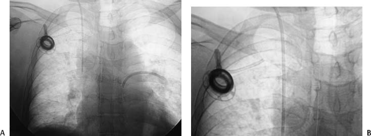
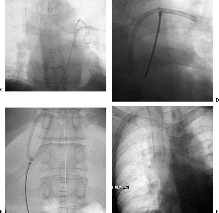
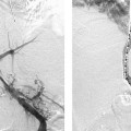
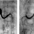
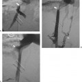
CASE 7 A 43-year-old female presented to an emergency department with “heart palpatations.” She had been treated 5 years earlier for Hodgkin’s disease and received a surgically placed chest port inserted via the subclavian vein. In the emergency department, the patient’s symptoms spontaneously resolved and a diagnosis of supraventricular tachycardia was presumptively made. No imaging was performed. One week later, symptoms recurred and the patient returned to the hospital. A chest radiograph was obtained. Figure 7-1 Catheter pinch-off with foreign body retrieval. (A,B) Scout view shows fractured catheter with fragment in the expected region of the left pulmonary artery. (C) Fluoroscopic image shows loop snare adjacent to catheter fragment. (D) Fluoroscopic image demonstrates fragment captured by snare. (E) Fluoroscopic image shows catheter being retracted to the common femoral vein via the inferior vena cava. (F) Final fluoroscopic image after removal of fragment and chest port. Scout view shows a left subclavian chest port with the catheter portion of the device fractured and embolized to the heart and pulmonary artery (Figs. 7-1A, B). Catheter pinch-off syndrome with fracture and embolization. The patient was brought to the interventional radiology suite and the right groin and left chest were prepped. A 7-French (F) sheath was inserted into the right common femoral vein and a loop snare was advanced from the groin to the heart (Fig. 7-1C
Clinical Presentation
Radiologic Studies
Diagnosis
Treatment
![]()
Stay updated, free articles. Join our Telegram channel

Full access? Get Clinical Tree


