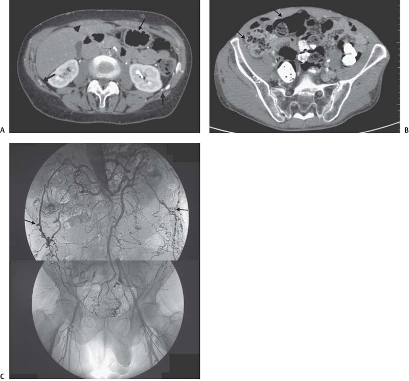
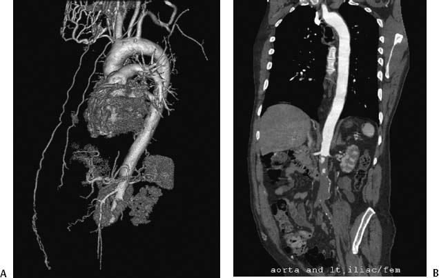
CASE 8 A 55-year-old male presented with complaints of buttock claudication and impotence. Physical exam revealed very weak femoral pulses. A computed tomographic angiogram (CTA) was requested. Figure 8-1 Leriche syndrome. (A) CT image shows all three routes of collateral bloodflow: posterior (large arrows), anterior (arrowheads), and middle (small arrow). (B) CT image shows right posterior deep circumflex iliac artery (large arrows) and right inferior epigastric artery (arrowheads) collateral routes cephalad to their connection with the right common femoral artery. (C) Digital subtraction angiogram shows infrarenal aortic occlusion. Note posterior (large arrows) and middle (small arrows) collateral routes of arterial blood flow. Anterior route of collateral flow will not be seen on abdominal aortogram because this route begins at the level of the aortic arch (internal mammary arteries). CTA shows complete occlusion of the aorta and common iliac arteries (Figs. 8-1A, B). Angiography confirms these findings (Fig. 8-1C). Aortic occlusion or Leriche syndrome. Aortobifemoral bypass graft.
Clinical Presentation
Radiologic Studies
Diagnosis
Treatment
Discussion
Background
Related posts:


Stay updated, free articles. Join our Telegram channel

Full access? Get Clinical Tree


