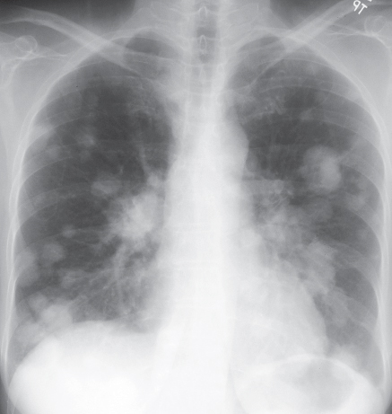
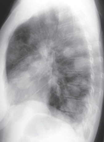
CASE 80 50-year-old woman status post hysterectomy for uterine cancer PA (Fig. 80.1) and lateral (Fig. 80.2) chest radiographs demonstrate multiple bilateral well-defined spherical pulmonary nodules and masses, which are most numerous in the lung bases. Pulmonary Metastases • Primary Pulmonary Lymphoma • Multicentric Lung Cancer • Hematogenous Infection • Vasculitis Fig. 80.1 Fig. 80.2 The term metastasis is generally defined as the transfer of disease from one organ to another that is not in direct contiguity. In the setting of neoplasia, metastases are characteristic of malignancy. Pulmonary metastases represent the most frequent pulmonary neoplasm. Depending on the location of the primary tumor, lung metastases may represent early or late neoplastic dissemination. Secondary lung neoplasia may occur via hematogenous, lymphatic, and tracheobronchial routes. Pulmonary metastases typically result from hematogenous dissemination of malignancy. The neoplastic cells are transported to the lung and “arrest” in its capillary bed. Thus, most primary neoplasms that produce lung metastases have a rich vascular supply and a venous drainage into the systemic circulation, with the lung as the filtering organ. Pulmonary metastases may in turn metastasize and produce disseminated malignancy. Patients with pulmonary metastases may be entirely asymptomatic. When metastases are numerous, large, and/or involve the airways and pleura, affected patients may experience dyspnea, cough, chest pain, and respiratory failure. Patients with lymphangitic carcinomatosis typically present with dyspnea. Patients with sarcoma metastatic to the lung may present with acute chest pain because of secondary spontaneous pneumothorax. Most patients with pulmonary metastases have a known history of malignancy. Rarely, patients present with pulmonary metastases from an unknown primary neoplasm.
 Clinical Presentation
 Radiologic Findings
 Diagnosis
 Differential Diagnosis


 Discussion
Background
Etiology
Clinical Findings
Imaging Findings
Chest Radiography
Stay updated, free articles. Join our Telegram channel

Full access? Get Clinical Tree


