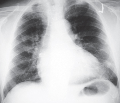
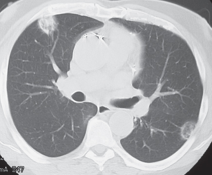
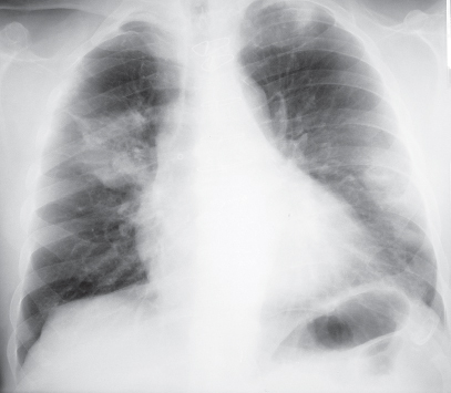
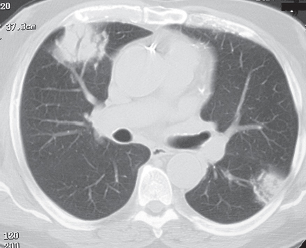
CASE 82 Asymptomatic 71-year-old man evaluated because of an abnormal chest radiograph PA chest radiograph (Fig. 82.1) demonstrates multi-focal bilateral nodular opacities without associated lymphadenopathy or pleural effusion. Unenhanced chest CT (lung window) (Fig. 82.2) shows multi-focal subpleural nodular opacities of heterogeneous attenuation. Biopsy confirmed the diagnosis of primary pulmonary lymphoma of mucosa-associated lymphoid tissue (MALT). PA chest radiograph (Fig. 82.3) obtained three years later demonstrates interval growth and coalescence of multi-focal nodular opacities. Unenhanced chest CT (lung window) (Fig. 82.4) confirms interval growth of pulmonary lesions, which exhibit internal air bronchograms. Primary Pulmonary Lymphoma; Marginal Zone B-Cell Lymphoma of MALT • Multicentric Lung Cancer • Pulmonary Metastases • Multi-focal Pneumonia • Cryptogenic Organizing Pneumonia Fig. 82.1 Fig. 82.2 Fig. 82.3 Fig. 82.4 Pulmonary lymphomas are subdivided into those lymphomas limited to the lung (primary) and those in which the lung is affected secondarily. A primary pulmonary lymphoma is defined as an extranodal pulmonary lymphoma without evidence of extrapulmonary involvement at diagnosis and during the subsequent three months. It is a rare lung malignancy and comprises approximately 0.5% of all primary lung neoplasms. Most (70–90%) primary pulmonary lymphomas are marginal zone lymphomas of MALT. Primary pulmonary diffuse large B-cell lymphomas account for approximately 5–20% of all primary pulmonary lymphomas. A secondary pulmonary lymphoma is much more frequent, occurs in approximately 50% of patients with lymphoma, and is more common in patients with Hodgkin lymphoma. Primary pulmonary lymphomas
 Clinical Presentation
 Radiologic Findings
 Diagnosis
 Differential Diagnosis
 Discussion
Background
Etiology
Related posts:
![]()
Stay updated, free articles. Join our Telegram channel

Full access? Get Clinical Tree


Radiology Key
Fastest Radiology Insight Engine


