Axial non-contrast CT scan through the lesion.
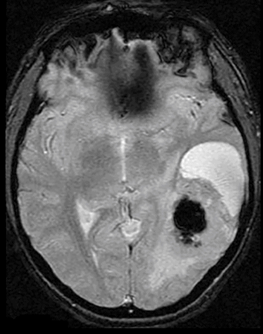
Axial GRE through the lesion.
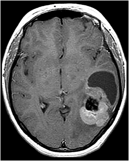
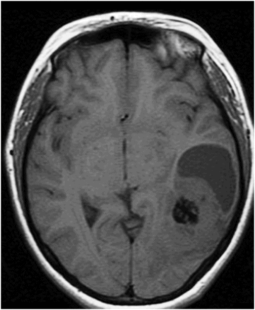
Axial T1WI with contrast through the lesion.
Astroblastoma
Primary Diagnosis
Astroblastoma
Differential Diagnoses
Supratentorial ependymoma
Pleomorphic xanthoastrocytoma
Ganglioglioma
Supratentorial juvenile pilocytic astrocytoma
Metastasis
Oligodendroglioma
Imaging Findings
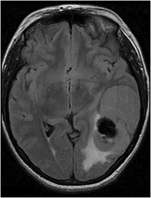
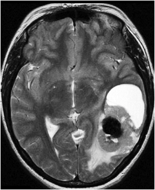
Fig. 85.1: Axial non-enhanced CT image of the brain demonstrated a complex, left temporal lobe mass with a centrally located gross calcified component, surrounded by non-calcified tumor tissue and an anterior polar cystic lesion. Fig. 85.2: Axial T1W image showed the complex and heterogeneous solid-cystic mass with a central, marked hypointense nodule, with adjacent cystic and solid components. Note that the calcified area appears hypointense. Fig. 85.3: Axial FLAIR image demonstrated mild peritumoral abnormal signal posterior to the mass. Note that there is signal void at the central calcified nodule. Fig. 85.4: Axial GRE image demonstrated signal void at the centrally located calcified nodule. Fig. 85.5: Axial T2W image demonstrated that the lesion itself has three major components: a centrally located hypointense (due to calcification) component; a surrounding, solid component demonstrating multiple, small cysts; and a larger cystic component at the anterior aspect. Fig. 85.6: Axial T1W image with contrast demonstrated enhancement of the soft tissue component (surrounding the calcified nodule), which presented moderate enhancement, with no cystic parietal enhancement, and no significant contrast uptake in the calcified nodule.
Discussion
Astroblastoma should be included in the differential diagnosis of a relatively benign-appearing solid-cystic tumor with calcification, multiple cysts in the solid component (bubbly appearance), and heterogeneous enhancement in a relatively young patient.
Many tumors can have similar appearances. Supratentorial ependymomas usually have a cystic component with high signal on T2-weighted images. Ependymomas may also present cystic component, which are usually simple with the solid component lacking a bubbly appearance as seen in cases of astroblastomas. Ependymomas tend to show moderate amount of peripheral T2 hyperintensity for their size due to prominent peritumoral infiltration invading the adjacent parenchyma. Pleomorphic xanthoastrocytoma, ganglioglioma, rare supratentorial juvenile pilocytic astrocytomas, and rare supratentorial hemangioblastomas all generally present with a strongly enhancing mural nodule within a single large cyst, a very distinct pattern seen in cases of astroblastomas. Oligodendrogliomas are also rare lesions that arise in the white matter and invade the cortex. They may be large, have cystic components, which may be seen as bubbly components, and have gross grouped calcification (present in 70–80% of the cases). Calcification is a consistent imaging feature seen in most reported cases of astroblastomas, which tend to be punctate.
Astroblastomas are distinctive neuroepithelial tumors of no definitive origin initially described by Bailey and Cushing in 1924 and further characterized by Bailey and Bucy in 1930. This is a rare (only 0.45–2.8% of all neuroglial tumors) tumor. Astroblastomas can occur in any age group, but generally occur in children and young adults, with no gender predilection. Clinical signs and symptoms depend on the localization and size of the neoplasm, with headache and seizures being the most frequent manifestations.
The origin of astroblastomas has been debated but a definitive precursor cell has not yet been identified. Recently, it has been postulated that astroblastomas may arise from abnormally persisting groups of embryonal precursor cells during embryogenesis such as tanycytes, a transitional cell type that is between astrocyte and ependymal cells.
Astroblastomas share features of astrocytomas, ependymomas, and of non-neuroepithelial tumors on histopathology owing to their astroblastic aspects. Lack of fibrillarity is an essential feature in distinguishing astroblastomas from other glial neoplasms. Immunohistochemically, astroblastomas are immunoreactive for glial fibrillary acidic protein (GFAP), S-100 protein, and vimentin. The majority of lesions display a focal cytoplasmic immunoreactivity for epithelial membrane antigen (EMA). Astroblastomas are defined histologically by the presence of perivascular pseudorosettes and prominent perivascular hyalinization.
Two distinct histologic types have been described: a low-grade type, in which a better-differentiated pattern was apparent and a favorable postoperative prognosis may be expected; and a high-grade type, showing more anaplastic microscopic features, in which postoperative survival is usually short. High-grade lesions show focal or multifocal regions of high cellularity, anaplastic nuclear features, elevated mitotic indices, vascular proliferation, and necrosis with pseudopalisading.
Although malignant astroblastomas may show infiltration of brain parenchyma, most of them are non-infiltrating lesions. Total resection is reported to be the method of treating an astroblastoma. Close follow-up of all cases and adjuvant therapy for high-grade and recurrent cases is recommended. The more favorable prognosis is almost invariably associated with circumscription of the tumor that might permit the total resection of tumor in all grades.
Imaging findings may also assist in the preoperative diagnosis of astroblastomas. Typically, they present as large, peripherally located supratentorial masses. On MRI, they are cystic and solid tumors with a bubbly appearance to the solid component. Disregarding their large size, astroblastomas have relatively little T2 adjacent hyperintensity, possibly because of their lack of tumor infiltration of the surrounding brain parenchyma. Astroblastomas also tend to demonstrate low signal on T2-weighted images and may demonstrate increased density on CT scans. Even though astroblastomas are classically described as non-infiltrating masses in the cerebral hemispheres, tumor may be seen in the corpus callosum, cerebellum, brainstem, and optic nerves. Astroblastomas typically show rim enhancement on CT and T1W MRI. However, heterogeneous gadolinium enhancement may be seen. Less common findings are intratumoral hemorrhage and intraventricular location.
Stay updated, free articles. Join our Telegram channel

Full access? Get Clinical Tree


