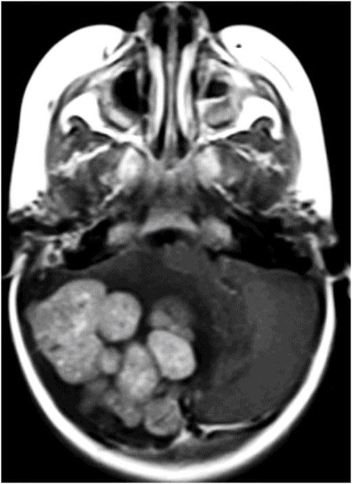
Axial T2WI through the posterior fossa.

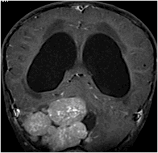
Coronal T1WI with contrast through the posterior fossa.
Medulloblastoma with Extensive Nodularity
Primary Diagnosis
Medulloblastoma with extensive nodularity
Differential Diagnoses
Atypical teratoid/rhabdoid tumor
Lhermitte-Duclos disease
Atypical choroid plexus carcinoma
Imaging Findings
Fig. 87.1: Axial T2WI image through the posterior fossa demonstrated an extensive nodular T2 hypo- to isointense mass in the right side of the posterior fossa involving both the vermis as well as the right cerebellar hemisphere, with extensive peritumoral abnormal T2 hyperintensity, severe mass effect, and leftward displacement of the fourth ventricle and brainstem. Fig. 87.2: Axial DWI image through the same level demonstrated increased signal within the nodular areas of the tumor. With low ADC values, this feature is suggestive of decreased diffusivity. Fig. 87.3: Axial T1WI with contrast through the same level demonstrates intense enhancement of the nodular components. Extensive nodularity of the lesion is better demonstrated on this image. Fig. 87.4: Coronal T1WI with contrast through the posterior fossa also demonstrated an intensely enhancing extensively nodular lesion involving the midline, as well as the right cerebellar hemisphere.
Discussion
Imaging demonstrated an extensively nodular tumor involving the vermis as well as the right cerebellar hemisphere, with extensive peritumoral T2 hyperintensity and significant mass effect to the fourth ventricle and the brainstem. The mass is iso- to hypointense on T2WI and demonstrates diffusion restriction, suggesting a highly cellular tumor. In a five-year-old patient, an aggressive-looking mass in the posterior fossa with high cellularity is almost diagnostic of medulloblastoma (MB). The extensive nodularity suggests medulloblastoma with extensive nodularity (MBEN).
Atypical teratoid/rhabdoid tumor can have a similar imaging appearance but it is extremely rare in the posterior fossa and in this age group. Predominantly a hemispheric lesion, Lhermitte-Duclos disease (LDD) does not demonstrate intense contrast enhancement or show decreased diffusivity. In fact, diffusivity is increased in LDD. Choroid plexus carcinoma also can have a similar appearance, but it is an extremely rare tumor. In contrast to MBEN, it predominantly involves the atria of the lateral ventricle, rather than the fourth ventricle.
Medulloblastoma is a high-grade (WHO grade IV) embryonal tumor of the cerebellum. It is the most common malignant brain tumor of childhood. It occurs at all ages with the peak incidence between four and seven years of age. Seventy percent of all MBs occur in children less than seven years of age. Overall 65% of all MBs are encountered in males. At least 75% of childhood MBs arise from the vermis, projecting into the fourth ventricle. Increased cerebellar hemisphere involvement is correlative with increasing patient age.
Typical MBEN presenting symptoms include cerebellar syndrome (truncal ataxia and disturbed gait) with features of increased intracranial pressure including headache and morning vomiting due to mass effect, as well as obstructive hydrocephalus.
Medulloblastoma with extensive nodularity is one of the known histopathologic subtypes of MB. Previously, MBEN was referred to as cerebellar neuroblastoma. This tumor usually occurs in infants and typically has an expanded lobular architecture. The extensive nodularity is thought to be due to the presence of excessive small cells with round nuclei in the background of the reticulin-free zone. With treatment, the tumor may dedifferentiate into a predominantly ganglion cell tumor.
Deeper understanding of the molecular/genetic pathways and correlative histopathology of MB has led to the currently accepted classification system that divides MB into four different subgroups. These four subgroups include the WNT group, SHH group, Group 3, and Group 4 (see Table 87.1); MBEN is seen only in the SHH subgroup of MBs.


Stay updated, free articles. Join our Telegram channel

Full access? Get Clinical Tree


