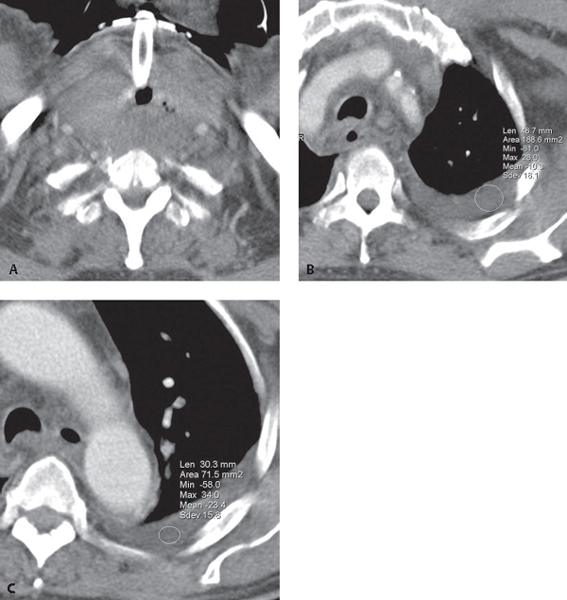
CASE 87 59-year-old man stabbed in Zone I (demarcated by thoracic inlet inferiorly and cricoid cartilage superiorly) and Zone II (mid-portion of neck from cricoid cartilage to angle of mandible) of the left neck six days earlier. Emergent tracheostomy was required. A left pleural effusion evolved on chest radiography (not illustrated) over the past several days. Contrast-enhanced CT at the T1 level (Fig. 87.1A) reveals a mid-line tracheostomy device and a large, eccentric, left-sided retrotracheal fluid collection. Note the lateral displacement of the great vessels and residual foci of pneumomediastinum. CT images at the branch vessel (Fig. 87.1B) and aortic pulmonary window level (Fig. 87.1C) show a low-attenuation left pleural effusion (–10 to –23 HU). Subsequent pleural fluid analysis showed a high concentration of emulsified fats and triglycerides (>110 mg/dL). Fig. 87.1 Chyloma with Chylothorax; Traumatic Thoracic Duct Injury None Chylothorax is the accumulation of chyle (i.e., lymphatic fluid) in the pleural space. Trauma is the second leading cause of chylothorax (25%). Thoracic duct disruption is complicated by accumulation of lymph in the extrapleural mediastinum, which forms a mass-like lesion called a chyloma. Subsequent rupture of the chyloma into the pleural space causes the chylothorax. The anatomic course of the thoracic duct and the site of injury determine the situs of the chylous effusion. The thoracic duct originates off the cisterna chyli at L2 and courses to the right of the spine ventral and medial to the azygos vein. Therefore, injury to the lower half
 Clinical Presentation
 Radiologic Findings

 Diagnosis
 Differential Diagnosis
 Discussion
Background
![]()
Stay updated, free articles. Join our Telegram channel

Full access? Get Clinical Tree


