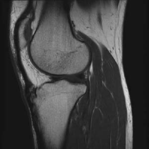
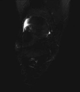
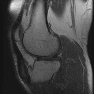
CASE 9 Anthony G. Ryan and Peter L. Munk A 13-year-old boy presented to his family doctor with a history of anterior knee pain. Direct questioning revealed he had recently taken part in a track and field competition. Physical exam revealed focal tenderness over the tibial tuberosity. He had full range of passive motion but a somewhat limited knee extension secondary to the pain. The practitioner sent the boy for a radiograph, which revealed no significant abnormality, but because the boy had ongoing pain, he was referred to an orthopedic surgeon, who requested an MRI. Figure 9A Figure 9B Figure 9C A sagittal T1 (Fig. 9A) image shows minimal fragmentation of the proximal aspect of the tibial tuberosity apophysis and fiber discontinuity in the adjacent patellar tendon insertion. A coronal short tau inversion recovery (STIR) sequence (Fig. 9B) through the tibial tuberosity reveals focal high signal intensity surrounding the cephalad fragment. Sagittal T2 (Fig. 9C) shows a markedly thickened patellar tendon at the level of its insertion with demonstrated high signal intensity within the tendon. Osgood-Schlatter disease. The etiology of Osgood-Schlatter disease remains unclear, although there is consensus that trauma is the underlying cause. The debate arises in how one describes the actual injury; that is, all the following may be described in the complex of injuries described as Osgood-Schlatter disease: a cartilaginous avulsion fracture of the tibial tuberosity, a tear of the patellar tendon at its tibial insertion, or chronic tendinitis of the patellar tendon with secondary ossification. The features common to all the above involve a tendon abnormality, with or without an attendant osseus component, as evinced by the characteristic findings of persistent ossific fragments within a thickened patellar tendon in the later stages of the disease. This begs the question regarding the existence of an actual fracture at the time of injury or whether it is ossification within a chronic tendinitis.
Osgood-Schlatter’s Disease
Clinical Presentation


Radiologic Findings
Diagnosis
Differential Diagnosis
Discussion
Background
Stay updated, free articles. Join our Telegram channel

Full access? Get Clinical Tree


