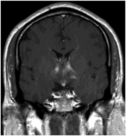
Coronal T2WI through the anterior commissure.

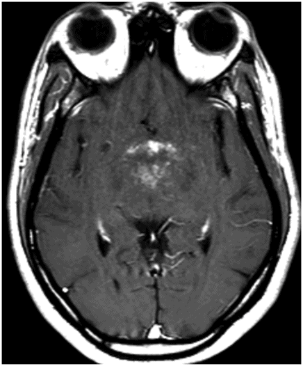
Postcontrast axial T1WI sequence through the level of the hypothalamus.
Anti-Ma2-Associated Paraneoplastic Encephalitis
Primary Diagnosis
Anti-Ma2-associated paraneoplastic encephalitis (AMAPE)
Differential Diagnoses
Limbic encephalitis due to other causes
Herpes encephalitis
Metabolic/toxic encephalopathy
Neurosyphilis
Imaging Findings
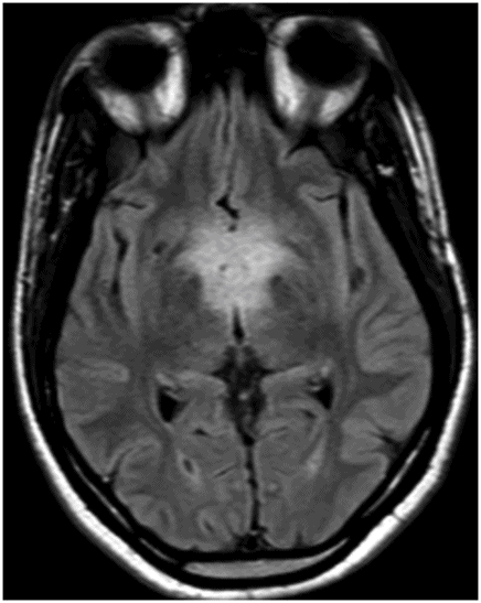
Fig. 96.1: Coronal T2WI through the anterior commissure demonstrates diffuse, confluent abnormal T2 signal involving the bilateral hypothalamus and anterior thalamus. Fig. 96.2: Axial FLAIR image through the hypothalamus demonstrates abnormal FLAIR signal and subtle swelling involving the bilateral hypothalamus and anterior thalamus. Fig. 96.3: Postcontrast axial T1WI sequence through the level of the hypothalamus demonstrates abnormal patchy enhancement involving the bilateral hypothalamic regions. Fig. 96.4: Postcontrast coronal T1WI sequence through the same level as Fig. 96.1 demonstrates patchy enhancement involving the bilateral hypothalamus and the adjacent brain tissue at the margin of the third ventricle in the areas of abnormal T2 signal.
Discussion
Subacute-onset narcolepsy, memory problems, and weight gain suggest the presence of mixed diencephalitis and limbic encephalitis. Confirmed by MRI, these symptoms are also highly suggestive of paraneoplastic diencephalitis/limbic encephalitis, which in a young male patient is usually due to AMAPE, secondary to testicular germ cell tumor. In this patient, testicular ultrasound demonstrated a 4 cm hard mass in his right testicle and bilateral testicular microlithiasis. Further CSF analysis was positive for anti-Ma2 antibody that confirmed the diagnosis.
A common manifestation of many paraneoplastic syndromes, limbic encephalitis has been characterized by the identification of multiple autoantibodies manifested in response to various systemic cancers. These include antibodies against 1) the N-methyl-D-aspartate receptor in ovarian teratoma, 2) the α-amino-3-hydroxy-5-methyl-4-isoxazolepropionic acid receptor in breast cancer and small cell lung cancer (SCLC), 3) the collapsin response mediator protein-5 in SCLC and thymoma, 4) the GABAB receptor in SCLC, 5) the antineuronal nuclear antibody type 1 (Hu) in SCLC, and of course, antibodies against Ma2 in testicular germ cell tumors. However, this patient only presented with testicular germ cell tumors and no other cancers.
Limbic encephalitis can be due to viral infection, especially herpes simplex virus in immune competent patients or HHV-6 infections in immunocompromised patients. However, this patient did not have any systemic symptoms of infection or any known immunodeficiency. The nature of subacute symptoms and negative CSF analysis for herpes viruses eliminated an infectious etiology. Neurosyphilis has no specific imaging abnormality and this patient did not have any history of congenital syphilis. Central nervous system manifestation of acquired syphilis within 25 years is extremely rare. Negative CSF FTA-ABS results further rule out the possibility of neurosyphilis. The patient was an otherwise healthy male, was not undergoing treatment for systemic disease, and he did not use recreational drugs, thus eliminating likelihood of metabolic/toxic causes of limbic encephalitis.
AMAPE usually occurs in young men with testicular germ cell tumors, most commonly non-seminomatous germ cell tumor. Other rare causes of AMAPE include breast cancers in women and non-small cell lung cancer in both older men and women. AMAPE most commonly presents with any combination of limbic encephalitis or diencephalitis and brainstem encephalitis in young male patients prior to the primary tumor diagnosis, in as many as 62% of cases. The association of AMAPE and testicular tumor is very strong. In fact, it is possible that young men with AMAPE will have a microscopic tumor below the detection threshold of imaging techniques. For this reason, both ultrasound and MRI should be performed to reveal potential microcalcification or subtle enhancement on MRI. Empiric orchiectomy has also been suggested if there is clinical deterioration, testicular enlargement, previous cryptorchidism, or evidence of testicular microlithiasis, in the absence of other tumors.
Neurologic manifestations of AMAPE include isolated limbic encephalitis, diencephalitis, or brainstem encephalitis, or any combination of these diseases. The most common presentation is brainstem encephalitis with either limbic encephalitis or diencephalitis. Seizure is another common manifestation of AMAPE. Common manifestations of limbic encephalitis include short-term memory deficits, confusional states, and decline of cognitive functions. Weight gain or loss, excessive sleepiness, narcolepsy-cataplexy, endocrinopathy, and hyperthermia have been described in cases of diencephalitis. Abnormal eye movements, opsoclonus, oculogyric crisis, Horner’s sign, dysarthria and dysphagia, facial weakness, and sensorineural hearing loss have been described as common presentation of brainstem encephalitis associated with AMAPE.
Although brainstem encephalitis with limbic encephalitis or diencephalitis combination is the most common clinical manifestation of AMAPE, the most common imaging abnormality is a combination of limbic encephalitis and diencephalitis. Abnormal T2 signal is seen in the mesial temporal lobe(s) with or without enhancement. This can be unilateral or bilateral. Similar imaging abnormalities are noted in the hypothalamus with varying extension to the basal ganglia and thalamus in diencephalitis. Abnormal T2 signal in the midbrain, pons, anterior medulla, and middle cerebellar peduncles has been described in association with brainstem encephalitis.
Anti-Ma2 antibody is seen in the CSF as well as in serum.
Stay updated, free articles. Join our Telegram channel

Full access? Get Clinical Tree


