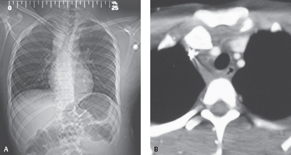
CASE 98 15-year-old male who fell off dirt bike while attempting to jump over an embankment AP CT scout (Fig. 98.1A) shows asymmetric alignment of the clavicles. The right clavicle lies lower than the left. A right paratracheal opacity is also present. Contrast-enhanced chest CT (mediastinal window) (Fig. 98.1B) reveals posterior displacement of the right sternoclavicular joint with the medial edge encroaching on the innominate artery and a right paratracheal hematoma. Posterior Dislocation of the Right Sternoclavicular Joint None Significant direct or indirect blunt force to the shoulder girdle can cause traumatic dislocation of the sternoclavicular joint (SCJ). Anterior sternoclavicular dislocations are much more common (9:1) and usually result from an indirect mechanism such as a blow to the anterior shoulder that rotates the shoulder backward. Posterior sternoclavicular dislocations are relatively rare, accounting for <1% of all dislocations. Traumatic forces that drive the shoulder forward can cause such posterior dislocations. The proximity of the dislocated medial clavicular head to critical structures in the thoracic inlet (e.g., aerodigestive tract, great vessels, brachial plexus, and apical pleural reflections) can result in serious concomitant injuries the exclusion of which should be part of the radiologist’s search pattern. Fig. 98.1
 Clinical Presentation
 Radiologic Findings
 Diagnosis
 Differential Diagnosis
 Discussion
Background

Clinical Findings
Stay updated, free articles. Join our Telegram channel

Full access? Get Clinical Tree


