Abdomen: Traumatic Emergencies


Abdomen: Traumatic Emergencies


Jorge A. Soto

Clark O. West

Amanda M. Jarolimek

John H. Harris Jr.


Multidetector computed tomography (MDCT) has become the principal diagnostic modality for evaluation of blunt abdominal trauma, replacing diagnostic peritoneal lavage (DPL) in most circumstances. Trauma centers designated by the American College of Surgeons as level I or level II are required to have computed tomography (CT) available at all times. Radiologists skilled in trauma imaging are valuable members of the trauma team, where they provide timely and expert interpretations of abdominal and pelvic CT. Many of the skills learned for general CT diagnosis apply to trauma patients, but a detailed understanding of the appearance of traumatic injuries, patterns of injury, the assessment of the hemodynamic status, and common image artifacts prepares the radiologist for excellence in trauma CT diagnosis.

Sonography has become popular for the initial assessment of blunt abdominal trauma in some centers, but turf battles over control of this modality have developed in a few centers. These differences aside, there is little doubt that sonographic detection of hemoperitoneum in a hemodynamically unstable patient affects the initial management of the patient. Sonography provides an exciting opportunity for emergency radiologists and ultrasound technologists to become integral members of the trauma team, responding to trauma alerts and participating in the initial assessment of trauma patients.


GENERAL CONSIDERATIONS


Injury Packages

Injuries sustained in motor vehicle collisions depend on multiple factors, including the size of the vehicle, the position of the victim in the vehicle, the type of accident (frontal impact, lateral impact, sideswipe, rear impact, or rollover), body habitus of the victim, and the use and type of restraint device.1 Knowledge of the common patterns of injury in the lower chest, abdomen, and pelvis will aid the emergency radiologist in identifying injuries and assessing the significance of borderline findings. In all of the injury patterns discussed later, associated scalp, face, cranial, aortic, and extremity injuries are encountered but will be omitted for the sake of brevity. Unrestrained drivers involved in a frontal impact are at risk for injuries in the midline of the body, including sternal and rib fractures, pulmonary contusion and laceration, cardiac injury, and aorta and branch artery injuries. In the abdomen, lacerations of the spleen and liver and rupture of the small bowel are also encountered. Fractures of the pelvis and posterior dislocation of the hip complete the package.1 Unrestrained drivers involved in a lateral impact on the left side of the vehicle sustain left-sided injuries, including left-sided rib fracture; left lung contusion and laceration; lacerations of the spleen, left kidney, or left lobe of the liver; and fractures of the
pelvis.1 Passengers in a vehicle struck on the right side are prone to right-sided injuries, including right rib fractures, right lung contusions and lacerations, lacerations of the right lobe of the liver and right kidney, and pelvic fractures. Passengers are less prone to thoracic injuries and more prone to abdominal injuries than are drivers.1


Systematic Approach to Trauma: Computed Tomography Interpretation

As in general abdominal imaging, an ordered approach to trauma CT results in an interpretation that is both thorough and efficient. The “every organ on every slice” approach involves systematically looking at all of the images of the liver, spleen, pancreas, and so forth, until a mental checklist has been completed. To a large extent, such a general approach is applicable to CT of the trauma patient, but several life-threatening injuries should be added to the general approach. The following checklist is inspired by Halvorsen2:


  • Life-threatening, trauma-specific


  • Hemoperitoneum


  • Pneumothorax (lung windows)


  • Pneumoperitoneum (lung windows)


  • Hemodynamic status


  • Active arterial contrast extravasation


  • Liver and right paracolic gutter


  • Spleen and left paracolic gutter


  • Upper abdominal organs, including duodenum and pancreas


  • Retroperitoneum, including adrenals, kidneys, inferior vena cava, and aorta


  • Bowel and mesentery


  • Pelvis


  • Muscles, including abdominal wall, psoas, iliacus, and buttocks


  • Bones (bone windows)


  • Lowest cut (thigh hematoma)

This approach places emphasis on immediate identification of life-threatening conditions requiring urgent treatment, followed by a detailed analysis designed to minimize missed injuries. The necessity of obtaining lung windows and bone windows, in addition to soft-tissue windows, must be emphasized. If a computer workstation is available, interpretation of multiple windows can be accomplished without printing multiple sets of films.


Computed Tomographic Signs in Blunt Abdominal Trauma

Jeffrey has defined several useful CT signs for assessing abdominal visceral injuries.3 They are summarized below.


Sentinel Clot Sign

Clotted blood adjacent to the site of injury is of higher attenuation (45 to 70 Hounsfield units [HU]) than un-clotted blood (30 to 45 HU), which flows away from the site of injury (Fig. 15.1). When the source of intraperitoneal hemorrhage is not evident, the location of the highest attenuation blood clot is a clue to the most likely source.


Water Density Fluid Collections

Fluid collections with attenuation near that of water (0 to 15 HU) originate from rupture of the gallbladder, urinary bladder, small bowel, and cisterna chyli. When fluid of this density is encountered in the peritoneal cavity without an identifiable source, the safest assumption is that an undiagnosed bowel injury is present.


Hemodynamic Status

The size of the inferior vena cava (IVC) and, in children and young adults, the size of the aorta give valuable morphologic information that correlates with the patient’s intravascular volume and cardiac output, as discussed later.


Active Arterial Contrast Extravasation

Termed “active arterial extravasation” by Jeffrey et al. and “vascular blush” by others, this sign represents arterial bleeding into extravascular tissues during the 1 to 3 minutes between the time of intravenous injection of contrast medium and the time the abdomen is imaged.4, 5, 6 The extravasated arterial contrast material must be distinguished from extravasated oral contrast material and from a pseudoaneurysm of a vessel within the injured organ. Vascular extravasation typically appears as a poorly marginated, highattenuation collection, often measuring over 100 HU, surrounded by a large hematoma. Extravasated gastrointestinal contrast material is not usually surrounded by a hematoma, and a pseudoaneurysm usually has a well-defined margin. Delayed scanning that is routinely done to evaluate the urinary tract can be extended to the area of active extravasation, allowing a qualitative assessment of the rapidity of hemorrhage.


Figure 15.1. Grade II splenic laceration with sentinel clot. A: At the upper pole of the spleen, the lateral surface is lacerated (solid arrow). A high-attenuation blood clot (C) floats in the nondependent portion of the perisplenic space—the sentinel clot sign. Low-attenuation fluid is present in the right perihepatic space (open arrows). The electrocardiographic electrode over the left anterior chest causes a moderate streak artifact. B: In the hepatorenal recess, hemoperitoneum (open arrows) is only slightly higher in attenuation than the renal cyst lateral to the left kidney (curved arrow). Intraperitoneal fluid in the hepatorenal recess typically wraps around the tip of the liver, distinguishing it from retroperitoneal fluid.

Jeffrey states that conservative management is generally not appropriate in the face of active arterial hemorrhage on CT, favoring urgent surgery or angiographic embolization.3


Staging Injuries for Nonoperative Management

Most blunt injuries to the liver, spleen, and kidneys can be managed nonoperatively. Most patients will stop bleeding with supportive care and others will respond to angiographic embolization, avoiding surgery.7, 8, 9 CT is required for successful nonoperative management of liver, spleen, or kidney injuries to detect and determine the extent of the injury and to exclude other intraperitoneal or retroperitoneal injuries that require urgent surgery.10, 11 Describing the extent of injuries is facilitated by the use of a standardized staging system. Several systems have been proposed, generally based on the premise that larger and deeper lacerations, larger hematomas, and devitalized tissue are signs of more severe injuries with a worse prognosis and are more likely to require surgical management.12, 13, 14, 15 Staging systems are necessary to standardize nomenclature and establish a single classification system for comparative studies. However, no staging system is sufficient to predict precisely which patients will require surgery based on anatomic information alone. Instead, anatomic stage is one factor in a more complex equation. The presence of active arterial extravasation on CT has recently been recognized as another important predictor of the need for surgery or angiographic embolization.16 Important clinical factors include the patient’s general medical condition, hemodynamic status, and need for ongoing fluid resuscitation or blood transfusion.7, 17

The American Association for the Surgery of Trauma (AAST) has developed a series of organ injury scales. The staging parameters were originally developed for surgical description of injuries, but they have been used successfully for CT staging. Although imperfect, the AAST scales remain as the standard of communication among the trauma surgical community, and are therefore presented in this chapter. The use of these criteria for radiology reporting promotes detailed analysis of injuries and unambiguous communication of results. Other staging systems, such as the one described by Taylor and colleagues (Table 15.1), have been described but are not used extensively in practice.18


DIAGNOSTIC IMAGING TECHNIQUE


Computed Tomography


Technique

MDCT technology is optimal for assessment of blunt abdominal trauma. Helical (single detector) CT is acceptable, but it does not offer all the
capabilities available with MDCT and is more prone to respiratory motion artifacts. Administration of intravenous contrast medium is mandatory to detect all possible injuries. Unenhanced CT is performed rarely in trauma, limited to patients with a well-documented severe allergy to contrast material. Evaluation of vessels and solid organs is not complete unless intravenous contrast material is administered. Patient preparation is very important but must be accomplished efficiently. To minimize streak artifacts, metal objects within the scanning field should be removed or repositioned, including metal buckles on patient restraints, belts, electrocardiography electrodes (Fig. 15.1) and connecting cables, and metal or metal-reinforced backboards (Fig. 15.2). Some backboards cause such severe artifacts that the patient should be transferred to a more “CT friendly” wooden or plastic backboard. Once the patient is on the CT table, the patient’s upper extremities should be raised above the head to avoid streak artifact. When upper extremity injuries preclude repositioning of the arms, placing the upper extremity close to the patient’s side and enlarging the scanning field to include the upper extremity reduces artifacts. Alert and cooperative patients should hold their breath during the imaging of the upper abdomen to minimize respiratory motion artifacts. However, successful breath holding is often not possible and scanning during shallow, quiet breathing is often necessary.


TABLE 15.1 Grading Criteria for Abdominal and Retroperitoneal Injury


Level 0—Negative


No manifestation of injury to intra-abdominal or retroperitoneal organs (injury to osseous structures was not included).


Level 1—Indeterminate


Findings that could be caused by incidental pathologic conditions rather than trauma-related injury.


Possibility that the finding is a normal anatomic structure or variant.


Abnormality present, may be related to artifact.


Abnormality may be such a subtle manifestation that it does not meet diagnostic criteria for definite injury.


Level 2—Minor


Minimal injury to intra-abdominal or retroperitoneal organs (e.g., small laceration, subcapsular hematoma, or contusion involving 25% or less of organ).


Minimal hemoperitoneum: small amount of fluid in two or fewer intraperitoneal spaces, with trace or no fluid in the third intraperitoneal space.


Small mesenteric hematoma only; no associated hemoperitoneum; no bowel thickening or enhancement.


Level 3—Moderate


Moderate liver and/or splenic laceration or minor injury with moderate hemoperitoneum (involving greater than 25% but less than 50% of the organ).


Mesenteric hematoma with minimal/moderate hemoperitoneum, but no bowel thickening or enhancement.


Retroperitoneal: renal injury, fracture with small perirenal or subcapsular hematoma, extravasation from collecting system, partial devascularization, adrenal hematoma.


Moderate hemoperitoneum: fluid present in more than trace amounts in three or more intraperitoneal spaces.


Level 4—Severe


Extensive liver injury of 50% or greater of the organ volume or less extensive injury but with large hemoperitoneum.


Extensive splenic laceration/fracture 50% or greater or involving hilum, with perisplenic hematoma and presence of any hemoperitoneum.


Bowel injury: thickening of bowel wall, enhancement of bowel, contrast medium extravasation from bowel, or free air.


Pancreatic fracture, or intrapancreatic or peripancreatic hematoma with hemoperitoneum.


Vascular contrast medium extravasation associated with any grade of solid organ injury.


Retroperitoneal injury: renal fracture with disruption, large perirenal hematoma, pedicle injury.


Large hemoperitoneum: fluid distending intraperitoneal spaces, fluid anterosuperior to the bladder.a


a Criterion for hemoperitoneum modified from Kane et al. From Taylor CR, Degutis L, Lange R, et al. Computed tomography in the initial evaluation of hemodynamically stable patients with blunt abdominal trauma: impact of severity of injury scale and technical factors on efficacy. J Trauma. 1998;44(5):893-901.


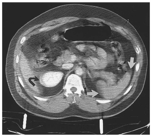
Figure 15.2. Streak artifact from backboard. At the splenic hilum and tip of the liver, severe streak artifact is so severe that the hemoperitoneum around the spleen is difficult to diagnose (straight arrows). The patient’s upper extremities at his side, the metal struts of the backboard, and respiratory motion cause the image degradation. Note the bright halo around the lateral portion of the right kidney, which simulates a subcapsular hematoma (curved arrow).

Intravenous catheters (18G or larger) should be placed in medially directed antecubital fossa or proximal forearm veins, so that these may be used for subsequent CT. Intravenous contrast (100 to 150 mL injected at a rate of 2 to 4 mL per second) is administered to all patients, and a 65- to 70-second delay is used for the portal venous phase acquisition. In addition, 5 to 7 minutes delayed images of the abdomen and pelvis are obtained selectively in those patients with injuries identified or suspected on the portal venous phase images.19, 20 Delayed scanning of the kidneys during this urographic phase of excretion of contrast material is essential to detect collecting system injuries. Delayed scans may also provide additional information about rapidly expanding hematomas and the rapidity of active extravasation of contrast material. Furthermore, delayed images help in characterizing extravascular collections of contrast-enhanced blood seen in the portal venous phase images. Importantly, these delayed phase images are acquired with a low radiation dose technique, reducing the tube current by 50% to 70% of that used for the portal venous phase series.19 Arterial phase images (25- to 30-second delay) may be added in severely traumatized patients.

Although all the earlier studies were based on CT scanning performed with oral contrast media,21, 22, 23, 24 the need for oral contrast in the setting of blunt abdominal trauma has been questioned by trauma surgeons, emergency medicine physicians, and radiologists.25, 26, 27 The main reasons are safety issues (risk for aspiration and subsequent complications), potential delay in diagnosis, and lack of proven substantial added diagnostic information for detection of significant bowel and mesenteric injuries. Extraluminal oral contrast is seen on CT in approximately 10% to 15% of patients with surgically proven significant bowel or mesenteric injuries.21, 22, 23, 24, 28 In addition to its low sensitivity, it is exceedingly rare to find extraluminal oral contrast as the only confirmatory sign of bowel or mesenteric trauma on CT. Almost 100% of the patients have additional highly suspicious CT findings that should alert the radiologist to the presence of a significant injury. Axial images reconstructed at both
1 to 1.25 mm and 3.75 to 5 mm section thicknesses are provided for interpretation. Orthogonal (coronal and sagittal) reformations are generated for all patients for all series acquired, both with 2.5-mm thickness and 2.5-mm reconstruction interval.


Diagnostic Performance of Computed Tomography

CT has been shown to be as accurate as DPL in detecting blunt abdominal injuries.29, 30 In a prospective comparison of CT and DPL, sensitivity, specificity, and accuracy were 97%, 95%, and 96% for conventional CT and 100%, 85%, and 95% for DPL.29 DPL involves placement of a catheter into the peritoneal cavity, aspiration of free fluid, instillation of normal saline, and aspiration of the lavage fluid. The fluid is analyzed for erythrocytes, white blood cells, amylase, bacteria, and bile, which are indicators of intraperitoneal injury. CT offers a number of advantages over DPL, including detailed anatomic evaluation of injuries, quantitation of associated hemorrhage, and detection of active arterial extravasation. CT staging is critical for successful nonoperative management of parenchymal organ injuries. CT excels at detection of retroperitoneal injuries, which are usually not detected by DPL or ultrasound. For these reason, CT has replaced DPL for evaluation of blunt trauma patients in most circumstances. In children, CT findings change the surgeon’s initial diagnosis in 84% of patients and result in changes in the patient management plan in 44%.31 Moreover, in a large, multi-institutional study of patients with suspected blunt abdominal trauma, CT had a negative predictive value of 99.63%.32 Based on their results, the authors of this study concluded that following a negative abdominal CT study using a modern technology, trauma patients could be safely discharged from the emergency department without a period of either inpatient or outpatient observation.32


Focused Assessment with Sonography for Trauma


Technique

Focused assessment with sonography for trauma (FAST) is a limited abdominal ultrasound examination performed early in the evaluation of patients with suspected blunt abdominal trauma. The examination should be completed within a few minutes and is designed primarily to search for peritoneal fluid. Although parenchymal organ injuries may be detected, the search for such injuries should not delay the examination. A six-point study is best performed with a 3.5 MHz sector transducer. The small footprint of the sector transducer is advantageous in establishing an acoustic window through intercostal spaces, a technique usually required for patients in distress who are unable to hold a deep breath for subcostal scanning. Longitudinal and transverse images should be recorded in the following areas: (a) the right subphrenic space above the liver, (b) the hepatorenal recess (Morrison pouch), (c) the left subphrenic space above the spleen, (d) the perisplenic space at the inferior margin of the spleen, (e) the peritoneal recess of the pelvis, and (f) the pericardium.33 Acoustic windows for this examination are usually found in the right midaxillary line either intercostally or subcostally, the left posterior axillary line either intercostally or subcostally, the midline 4 cm superior to the symphysis pubis, and the subxiphoid region.

Unclotted intraperitoneal blood is hypoechoic, typically with low-level internal echoes (Fig. 15.3). Clotted blood may be either isoechoic or hyperechoic and may be indistinguishable from the adjacent parenchyma of the liver or spleen. Failure to visualize a normal spleen is a sign that the spleen is injured. One must not make the erroneous mental leap from “I don’t see much around the spleen,” to “Therefore, nothing much is wrong with the spleen.”

Advocates of FAST emphasize its ease of use and repeatability.29 Indeed, to avoid missing injuries, FAST may need to be repeated serially, but literature demonstrating the value of this approach does not yet exist.


Diagnostic Performance of FAST

In a prospective study of severely injured patients, FAST had a sensitivity of 86%, a specificity of 99%, and an accuracy of 98% for the detection of abdominal injuries requiring surgery.34 However, false-negative examinations were encountered with liver and spleen injuries, particularly those not associated with hemoperitoneum. Chiu and colleagues reported that of 52 abdominal injuries evaluated by both FAST and CT, 15 (29%) were evaluated as negative by FAST but were shown to include injuries to the liver or spleen without hemoperitoneum by CT.35 Three patients required laparotomy to control bleeding. These data illustrate that a negative FAST should be interpreted as indicating the absence of hemoperitoneum, not the absence of intra-abdominal injury.35


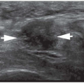
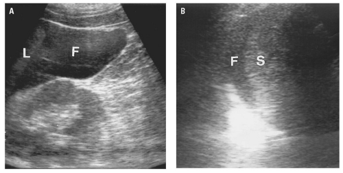
Figure 15.3. Hemoperitoneum. A: Longitudinal ultrasound image of the right upper quadrant obtained with a 3.5 MHz curved-array transducer demonstrates fluid (F) with low-level internal echoes in the hepatorenal recess adjacent to the tip of the liver (L). B: In a different patient, echogenic fluid (F) is present between the diaphragm and the upper pole of the spleen (S).

Because of its relatively low sensitivity, FAST should not be viewed as an equivalent replacement for either CT or DPL. A negative FAST should be viewed with some suspicion if the finding is not commensurate with the patient’s clinical presentation. On the other hand, a positive FAST does not necessarily mandate laparotomy. Hemodynamically stable patients with a positive FAST should have their injuries staged with CT, affording them the benefit of nonoperative management whenever possible. FAST is not reliable for assessment of the retroperitoneum; CT performs far better in this role and is indicated when injury mechanism, injury patterns, or the presence of hematuria suggest a retroperitoneal injury.


Figure 15.4. Pneumoperitoneum. In the midabdomen, a collection of free intraperitoneal air separates the parietal peritoneum of the anterior abdominal wall from the visceral peritoneum of the greater omentum (straight arrows). Air within an adjacent jejunal loop (curved arrow) surrounds thickened valvulae conniventes, which clearly establishes its intraluminal location.


BLUNT TRAUMATIC CONDITIONS


Peritoneal Cavity


Pneumoperitoneum

Pneumoperitoneum may be detected in the midabdomen beneath the anterior parietal peritoneum (Fig. 15.4) or in the upper abdomen over the anterior surface of the left lobe of the liver.
Bubbles of free air may be trapped between leaves of mesentery or in the peritoneal recesses. Lung windows are valuable in detecting pneumoperitoneum, which might be overlooked on soft tissue windows because free air may not be distinguishable from adjacent bowel gas at narrow window widths.

In trauma, extraluminal gas is a highly suggestive, but not pathognomonic, sign of bowel perforation.36, 37 Pneumoperitoneum is found on CT in 20% to 75% of patients with proven bowel perforations.27, 37, 38, 39, 40, 41, 42, 43, 44 The amount of free intraperitoneal gas varies widely and can be massive, filling all peritoneal compartments, or very small, with only a few bubbles noted outside of the bowel lumen. In all blunt trauma patients with any CT finding that could potentially be associated with a hollow viscus injury, images should be reviewed with lung or bone window settings, in addition to the routine soft tissue settings. This approach facilitates the detection of small extraluminal gas collections. In addition, it is important to look for pneumoperitoneum in both the portal venous phase and delayed phase images because, occasionally, the pneumoperitoneum may appear only on the second acquisition. Many patients with surgically proven perforations do not have any evidence of pneumoperitoneum on CT. Various reasons may explain this circumstance: the perforation may be contained or may partially seal spontaneously, developing ileus may prevent passage of gas into the abdominal cavity, or small gas collections may rapidly be reabsorbed through the peritoneal lining. A few potential causes of a false-positive finding of pneumoperitoneum should also be considered and be ruled out before the diagnosis of bowel perforation is made on the basis of free intraperitoneal gas alone. Causes of pneumoperitoneum without bowel trauma include intraperitoneal rupture of the urinary bladder with an indwelling Foley catheter, massive pneumothorax (especially if there is a coexistent diaphragmatic rupture), barotrauma, benign pneumoperitoneum (e.g., as observed in some patients with systemic sclerosis), and the occasional DPL. Pseudopneumoperitoneum is another potential cause of a false-positive diagnosis of free intraperitoneal gas and bowel rupture. Pseudopneumoperitoneum— air confined between the inner layer of the abdominal wall and the parietal peritoneum—may be found in patients who suffer injuries to the extraperitoneal segments of the rectum, rib fractures, pneumothorax, or pneumomediastinum, with collections of extraluminal gas accumulating between the deep layers of the abdominal wall and the parietal peritoneum.37 On CT, the appearance may closely resemble true pneumoperitoneum (Fig. 15.5). However, most patients with true pneumoperitoneum have collections of gas located deeper in the abdomen, often adjacent to the ruptured viscus, at the porta hepatis, or outlining the falciform ligament. If in doubt, delayed images or a decubitus series may help make this distinction.


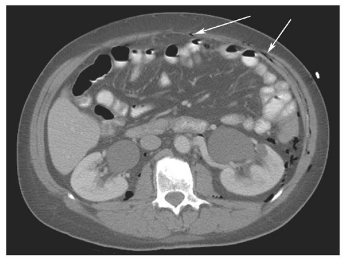
Figure 15.5. 58-year-old female patient with a pelvic fracture and a rectal wall tear, allowing leakage of gas into the extraperitoneal space of the pelvis. The axial CT image demonstrates gas in the retroperitoneum (surrounding the kidneys and descending colon) as well as in the extraperitoneal space of the anterior abdominal wall (“pseudopneumoperitoneum,” arrows). The patient underwent repair of the rectal tear, but no laparotomy was performed.


Peritoneal Fluid

Blood collects in the peritoneal cavity following injuries to the liver, spleen, bowel, and mesentery. As discussed under General Considerations, a high-attenuation “sentinel” clot may be seen near the site of bleeding, with lower attenuation blood elsewhere in the peritoneal cavity. In the supine position, blood from the liver collects in the hepatorenal recess and travels down the right paracolic gutter into the pelvis. From the spleen, blood passes along the phrenocolic ligament to the left paracolic gutter and the pelvis (Fig. 15.6).45 The volume of
hemoperitoneum may be estimated by searching for fluid in the perisplenic space, perihepatic space, hepatorenal recess, right and left paracolic gutters, and pelvis.46 Small collections of fluid are confined to one space; moderate collections are seen in two or more spaces; and large collections involve all spaces. Although the volume of hemoperitoneum will have an effect on patient management decisions, a large hemoperitoneum does not mandate laparotomy.10, 17


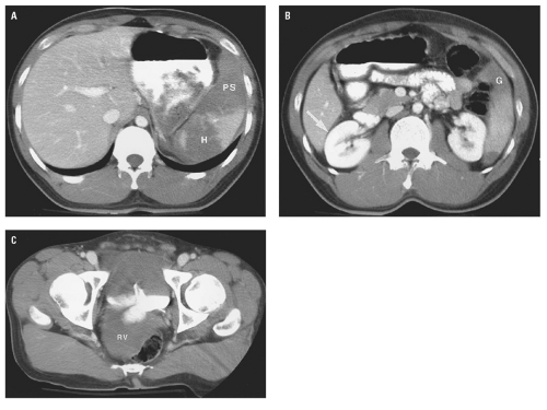
Figure 15.6. Grade IV splenic laceration with left-sided hemoperitoneum. A: Above the splenic hilum, the spleen is fragmented. Intraparenchymal hematoma (H) separates the fragments. Hemoperitoneum distends the perisplenic space (PS). B: At the lower pole, the hemoperitoneum enters the upper portion of the left paracolic gutter (G), but no hemoperitoneum is present in the hepatorenal recess (arrow). C: In the pelvis, hemoperitoneum fills the rectovesical space (RV).

Hemoperitoneum typically has an attenuation value of 45 HU or greater. However, in a retrospective analysis of 42 patients, Levine and colleagues reported that 16% of patients with liver and spleen injuries had hemoperitoneum with attenuation values greater than 45 HU, 60% had hemoperitoneum with attenuation values between 20 and 45 HU, and 24% had hemoperitoneum with attenuation values less than 20 HU.47 Thus, in some patients, hemoperitoneum may not be distinguishable from ascites, extravasated small intestinal fluid from bowel perforation, or intraperitoneal urine from bladder rupture.

The finding on CT of free intraperitoneal fluid in blunt trauma patients with an absence of identifiable injury causes difficulty in interpretation for both radiologists and trauma surgeons, particularly in males. In reproductive age female patients, isolated free fluid in the pelvis can be explained by ruptured ovarian follicular cysts. In males, the question is often raised as to what is the clinical importance of this finding; that is, whether a change in management should result from the presence of free fluid alone. In the late 1990s, research suggested that the finding of free intraperitoneal fluid in the setting of blunt trauma, with the absence of identifiable injury to explain the finding, necessitated exploratory laparotomy.48, 49 More recent work has led to a more conservative approach, with some of these patients being admitted for observation without immediate surgical intervention.32, 50, 51, 52, 53 With advances in CT technology, the sensitivity
for detecting small amounts of free intraperitoneal fluid has improved, and the radiologist must use all the tools available to help guide further management.

Unexplained hemoperitoneum raises the likelihood of an underlying small bowel or mesenteric injury.54 The attenuation of free blood in the peritoneal cavity is high (greater than 30 to 40 HU), as discussed previously. Drasin and colleagues showed that measuring the attenuation of the pocket(s) of free fluid may aid the radiologist and surgeon in determining the significance of the isolated free fluid in male patients.55 In their study, isolated free fluid with low attenuation was found in 2.8% of male patients who suffered blunt trauma, and none was proven to have a surgically important bowel or mesenteric injury.55 The mean attenuation of the pockets of fluid on the portal venous phase images was 13.1 HU (Fig. 15.7), considerably lower than the mean attenuation of free fluid in the group of patients who did have an identifiable injury to explain the finding on CT (mean attenuation 45.6 HU). Similar results were subsequently reported by Yu and colleagues, who found that 4.8% of male blunt trauma patients may have a small amount of free fluid in the pelvis as the only finding on CT.56 In this setting, the radiologist must carefully evaluate the CT images for findings that could explain the presence of low-attenuation fluid, such as intraperitoneal bladder rupture (urine) or biliary tract injury (bile). A trend for increased incidence of isolated free fluid can be seen in patients who receive higher volumes of fluid resuscitation.61 Also, the radiologist plays an important role by reviewing the images from the trauma scan at the CT scanner, and if a suspicious finding (such as free peritoneal fluid) is noted on the initial portal venous phase images, the decision to obtain delayed images should be made.


Figure 15.7. Isolated low attenuation free fluid. Coronal CT reformation demonstrates a small amount of low attenuation fluid (measured at 8 HU) in the pelvis (arrow). No additional findings to indicate presence of a solid organ or hollow viscus injury were present. The patient was treated conservatively and had an uneventful hospital stay.


Hemodynamic Status

CT provides an anatomic view of the patient’s hemodynamic status, but the signs can be nonspecific and must be interpreted with knowledge of the patient’s blood pressure, volume of resuscitation before CT, and estimated blood loss. A collapsed infrahepatic IVC on three or more consecutive images is a sign of intravascular volume depletion in a trauma patient who has received the usual 2 or more liters of crystalloid before CT scanning.57 Flattening of both renal veins frequently accompanies the collapsed IVC. In children with shock, a hypoperfusion complex has been defined: diffuse dilatation of the intestine with fluid; abnormally intense contrast enhancement of the bowel wall, mesentery, kidneys, aorta, and IVC; and reduced diameter of the abdominal aorta and IVC (Fig. 15.8). The hypoperfusion complex in children is associated with a high mortality rate. A similar hypoperfusion complex has been described in adults but has a less grave prognosis.58

Conversely, distention of the IVC with the absence of the normal image-to-image respiratory variation in IVC caliber may be an indicator of intravascular volume expansion associated with rapid resuscitation.59

The size of the IVC is affected by many factors, including hemodynamic status, IVC obstruction from thrombus or extrinsic masses, developmental
anomalies, or portosystemic shunting. Flattening of the IVC is encountered in outpatients undergoing CT for nontrauma reasons.60 However, in a hemorrhaging trauma patient, a flattened IVC indicates inadequate volume resuscitation. IVC flattening may precede clinical signs of shock. Recognition of intravascular volume depletion on CT should prompt aggressive volume resuscitation before the patient develops more overt signs of cardiovascular collapse.


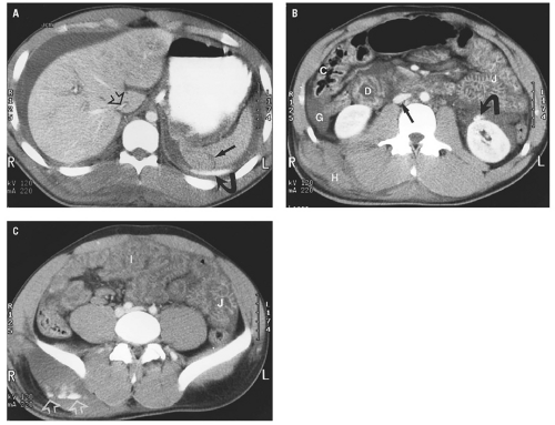
Figure 15.8. Splenic laceration with the hypoperfusion complex and active arterial extravasation in an 18-month-old infant. A: Image through the spleen demonstrates a grade II splenic laceration (straight arrow) with adjacent active arterial extravasation (curved arrow) and abundant hemoperitoneum surrounding the liver and spleen. Note the slit-like IVC (open arrow) as it passes through the liver. B: In the midabdomen, the duodenum (D), jejunum (J), and ascending colon (C) all have thickened walls and abnormally enhancing mucosa. Hemoperitoneum is seen in the right paracolic gutter (G). Active arterial extravasation from the anterior surface of the left kidney (curved arrow) results in a perinephric hematoma. The IVC (straight arrow) is flattened and the adjacent aorta is relatively small. A large hematoma (H) is present in the right flank. C: In the upper pelvis, abnormal mucosal enhancement is noted in the jejunum (J) and, to a lesser degree, the ileum (I). Active arterial extravasation is seen in the right buttocks (open arrows). (Case courtesy of Steven Ashlock, MD)


Liver and Biliary Tract


Liver

The liver is the second most frequently injured solid organ, after the spleen. In our experience, blunt liver injuries occur slightly less frequently than splenic injuries. The widespread use of MDCT allows recognition of minor liver injuries that were not recognized in the past.61 The right lobe is much more frequently injured than the left, with the
posterior segment of the right lobe most frequently injured.62 Most liver injuries cause hemoperitoneum, but approximately one-fifth do not.63 Liver injuries that do not produce hemoperitoneum will usually be missed by FAST, except in the unusual circumstance that a parenchymal laceration is seen directly by ultrasound. Liver injuries presenting without hemoperitoneum include minor liver injuries that do not cause significant bleeding, injuries that cause purely intraparenchymal hematoma, and injuries that disrupt the surface of the bare area of the liver and cause retroperitoneal hemorrhage.64


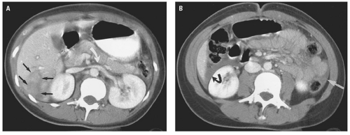
Figure 15.9. Grade III laceration of segments V and VI and complete pancreatic body laceration. A: Image through the right lobe of the liver shows a deep laceration extending to a portal vein branch in segment VI (straight black arrows). A hairline fracture is seen through the entire thickness of the pancreatic body (straight white arrows). Fluid between the pancreas and splenic vein (curved white arrow) indicates that this is a true pancreatic injury rather than a streak artifact over the pancreas. B: At the hepatorenal recess, hemoperitoneum (curved black arrow) surrounds the tip of the liver. Note a small amount of blood in the left paracolic gutter (white arrow).

Based on their morphologic appearance, injuries to the liver parenchyma may be classified into descriptive categories: laceration, fracture, intraparenchymal hematoma, contusion, and subcapsular hematoma.

Lacerations are the most common type of liver injury.62 Against a background of normally enhancing liver parenchyma, lacerations appear as linear or branching low-attenuation areas with sharp or jagged margins (Figs. 15.9, 15.10, 15.11 and 15.12).65 Lacerations frequently travel along vascular planes (portal vein branches and hepatic veins) and fissures (for the ligamentum teres and ligamentum venosum), which are anatomic weak zones. Multiple parallel lacerations may be described as “bear claw” lacerations. The depth of laceration and proximity to major vascular and biliary structures should be included in the radiology report. In particular, perihilar lacerations involving the first two or three divisions of portal venous branches are more likely
to be associated with biliary tract injuries.66 Lacerations extending to the proximal hepatic veins are important to identify because repair of hepatic vein injuries is technically difficult.67


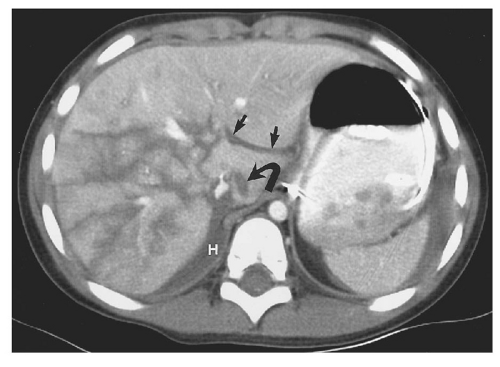
Figure 15.10. Grade V laceration of the entire right lobe of the liver. Multiple lacerations in a stellate pattern involve the entire right lobe of the liver, including the proximal portal vein branches and the intrahepatic IVC. Note retroperitoneal hematoma between the liver and the right hemidiaphragm (H) caused by lacerations involving the bare area. The fissure for the ligamentum venosum is filled with blood (straight arrows). Laceration in the caudate lobe is an unusual finding (curved arrow).


Figure 15.11. “Bear claw” liver laceration. Complex lacerations involving the left and right hepatic lobes, and extending to the porta hepatis.

The term “fracture” of the liver describes a through-and-through parenchymal laceration that may avulse a portion of the liver.

Intraparenchymal hematoma is a round or oval collection of blood retained within a liver laceration. A hepatic pseudoaneurysm or a focus of active arterial extravasation may be seen in the center of an intraparenchymal hematoma.


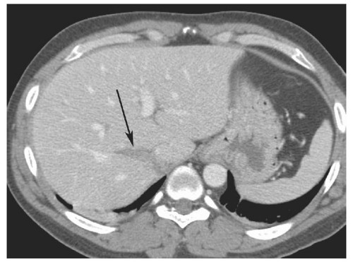
Figure 15.12. Grade II liver laceration (arrow) without associated hemoperitoneum.


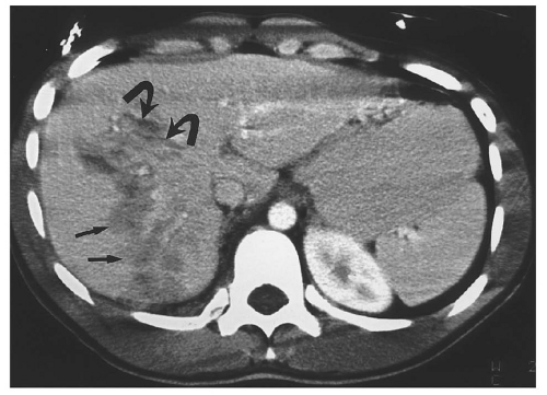
Figure 15.13. Hepatic contusion. The poorly defined area of low attenuation in the right lobe represents hepatic contusion (straight arrows). More well-defined lacerations are seen anteriorly (curved arrows).

Hepatic contusion is an area of minimal intraparenchymal hemorrhage that appears as a lowattenuation zone without well-defined laceration (Fig. 15.13).

A subcapsular hematoma is a lentiform collection of blood beneath an intact liver capsule that deforms the contour of the liver. Most subcapsular hematomas are located along the anterolateral aspect of the right lobe (Fig. 15.14).65 Respiratory motion artifact simulates the appearance of a subcapsular hematoma, as discussed later.


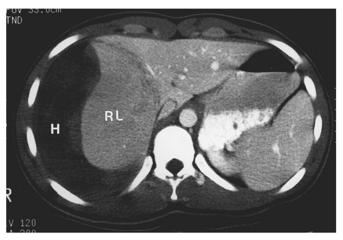
Figure 15.14. Large subcapsular hematoma of the liver. Above the porta hepatis, a large subcapsular hematoma (H) deforms the right lobe of the liver (RL). Note that the right lobe has diminished parenchymal enhancement compared to the left, probably due to the pressure exerted by the hematoma. (Case courtesy of Steven Ashlock, MD)


Periportal low attenuation is often the result of hemorrhage along the portal vein. On occasion, it is the only sign of liver injury.68 However, vigorous intravenous fluid administration or elevated central venous pressure—from tension pneumothorax or pericardial tamponade—may cause periportal low attenuation without liver injury.59

The detection of active arterial extravasation into the peritoneal cavity or within an intraparenchymal hematoma identifies patients who may need urgent endovascular or surgical treatment (Figs. 15.15 and 15.16). As with all extravascular collections of contrast-enhanced blood in trauma, the combined use of portal venous and delayed phase images is useful for characterizing contained lesions or true active extravasation.

Several pitfalls may complicate the interpretation of CT for liver injury. Respiratory motion artifact causes an indistinct gray margin around the right lobe of the liver, generally paralleling the contour of the liver (see Fig. 15.29). This appearance should not be confused with a subcapsular hematoma, which typically deforms the liver parenchyma. Respiratory motion artifact is the most common cause of false-positive diagnoses in abdominal trauma imaging.


Figure 15.15. Grade III-IV laceration of the right lobe with active arterial extravasation. Above the porta hepatis, a deep laceration involving segment VII has a focus of active hemorrhage at its base (curved arrow). Note the jet of active arterial extravasation extending to the periphery (straight arrows). Relatively dense blood is seen adjacent to the liver—the sentinel clot sign (C).


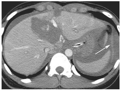
Figure 15.16. Massive liver laceration involving the right and left lobes, with large foci of intraparenchymal active extravasation as well as extravasation into the perisplenic space (arrow). Note also abundant hemoperitoneum.

Unopacified hepatic veins may be mistaken for a liver laceration (Fig. 15.17). This pitfall occurs when scanning commences early in the contrast bolus, before the hepatic veins are opacified. A focal area of fat—frequently in proximity to the ligamentum venosum—should not be confused with a hepatic laceration.69

Image artifacts may simulate lacerations. Most problematic are beam-hardening artifacts from adjacent ribs and streak artifacts from air-contrast interfaces, electrocardiographic electrodes, or the upper extremities within the imaging field.


Figure 15.17. Unopacified middle hepatic vein simulating a liver laceration. This image was made in the early vascular phase before there was filling of the portal or hepatic veins. The middle hepatic vein (arrows) could be mistaken for a deep laceration.


TABLE 15.2 American Association for the Surgery of Trauma Liver Injury Scale (1994 revision)


Only gold members can continue reading. Log In or Register to continue

Stay updated, free articles. Join our Telegram channel

Jun 20, 2016 | Posted by in GENERAL RADIOLOGY | Comments Off on Abdomen: Traumatic Emergencies

Full access? Get Clinical Tree

Get Clinical Tree app for offline access

Gradea


Injury description


I


Hematoma


Subcapsular, <10% surface area


Laceration


Capsular tear, <1 cm parenchymal depth


II


Hematoma


Subcapsular, 10%-50% surface area


Intraparenchymal, <10 cm in diameter


Laceration


1-3 cm parenchymal depth, <10 cm in length