Fig. 7.1
Anatomy of the superior abdominal cavity. The lesser sac is highlighted. K kidney, L liver, P pancreas, S spleen, St stomach
The transverse mesocolon, adjacent to the greater omentum—they in fact share the entrance into the transverse colon—connects the transverse colon to the retroperitoneum, along the anteroinferior margin of the pancreas, and contains the middle colic vessels.
The mesentery, in the small intestine, is a layer that connects the peritoneum to the posterior abdominal wall and contains the superior mesenteric artery and vein, lymph vessels and lymph nodes (Fig. 7.2). During its oblique course, of approximately 15 cm, from the duodenojejunal flexure to the ileocaecal region, it meets the head of the pancreas, the distal portion of the duodenum, the inferior vena cava, the abdominal aorta, the ureter and the right psoas muscle.
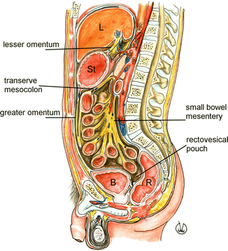
Fig. 7.2
Anatomy of the abdominal cavity, sagittal view. The parietal peritoneum layers are highlighted. B urinary bladder, L liver, R rectum, St stomach
The mesosigmoid connects the sigmoid flexure to the posterior abdominal wall and contains arterial and haemorrhoidal vessels.
In the pelvis, the broad ligaments, enveloping the Fallopian tubes, ovaries and uterine vessels, extend from the uterus to the lateral walls of the pelvis. The round ligaments serve to maintain the anteversion of the uterus. They contain lymph vessels, flow next to the inferior epigastric vessels, enter into the inguinal canals and continue towards the labia majora, where their fibres spread and mix the tissue of the mons pubis. The lateral, middle and median umbilical ligaments consist of peritoneal reflections, respectively, on the inferior epigastric vessels, umbilical arteries and urachus (the latter is an embryonic remnant going from the dome of the bladder to the umbilicus).
The above described folds and ligaments of the visceral peritoneum divide the peritoneal cavity into different parts; the main subdivision is caused by the transverse mesocolon, which separates the supramesocolic space from the inframesocolic one. The first one is between the diaphragm and the transverse mesocolon, and it is divided into the subphrenic and the subhepatic spaces, on the right, and subphrenic space and lesser sac, on the left. The right subphrenic space is separated from the left one by the falciform ligament which connects the anterior face of the liver to the anterior abdominal wall and that contains the round ligament (remnant of the umbilical cord). The left and right triangular ligaments originate at end of the reflections of the visceral peritoneum on the posterior face of the liver (coronary ligaments); the first of them connects the left hepatic lobe to the diaphragm, while the other one (also connecting the liver to the diaphragm) divides the perihepatic space into the subphrenic and the subhepatic spaces. The latter communicates, anteriorly, with the lesser sac, through the foramen of Winslow, and posteriorly, it forms the Morison’s pouch or hepatorenal recess. Between the reflections of the left and right coronary ligament, there is a hepatic region (so-called bare area of the liver) not covered with peritoneum, adjacent to the anterior pararenal space on the right. The peritoneal cavity has other similar “uncovered” areas where the abdominal wall and the viscera are in direct contact (which, such as the ascending and descending colon, are therefore in subperitoneal position) and where the passage between parietal and visceral peritoneal layers takes place. The subphrenic space on the left surrounds the spleen and it is delimited, inferiorly, by the phrenicocolic ligament which connects the left colic flexure to the left hemidiaphragm.
The inframesocolic compartment is divided into the right and left paracolic gutters, lateral paravesical recesses, vesicouterine pouch and rectouterine pouch, or pouch of Douglas, in women, while in men, into the rectovesical recess.
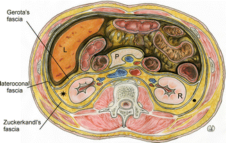
The retroperitoneum is the anatomical space behind the peritoneal cavity, delimited, anteriorly, by the posterior parietal peritoneum and, posteriorly, by the transversalis fascia covering the psoas and lumbar quadrate muscles. The retroperitoneum contains portions of colon, duodenum, pancreas, kidneys, ureters, suprarenal glands, abdominal aorta and inferior vena cava. It is usually divided into three spaces: posterior pararenal space, which contains fat only; perirenal space, delimited by the Gerota’s fascia, anteriorly, and the Zuckerkandl’s fascia, posteriorly, and containing kidneys, renal pelvis and proximal ureter, suprarenal glands and fat; and anterior pararenal space containing some segments of the colon, duodenum, pancreas and the root of the small bowel mesentery (Fig. 7.3). The lateral fusion of Gerota’s and Zuckerkandl’s renal fasciae at the level of the lateroconal fascia demarcates the perirenal fat from the posterior pararenal fat laterally. Superiorly, the renal fasciae merge together and are attached to the diaphragm. Caudally, the renal fasciae do not fuse together, and at the level of iliac crest, the anterior and posterior spaces are in potential communication. At this same level, the lateroconal fascia disappears as a distinct boundary so that the anterior pararenal space communicates laterally with the properitoneal fat of the flank stripe. In the pelvis, we find a single retroperitoneal space.