Fig. 9.1
Anatomy of the liver, frontal view. The surface is convex, asymmetrically divided into the left and right lobe by the falciform ligament, to which the round ligament (arrowhead) is attached. The gallbladder is highlighted in green
The anterosuperior surface is smooth and convex, almost completely adjacent to the diaphragm; the inferior or visceral surface, instead, is slightly concave and touches the underlying organs: there are three sulci or fissure, two longitudinal ones (right and left sagittal fissure) and a horizontal one, the transverse fissure. The latter represents the hilum, where we find the structures of the pedicle: portal vein and hepatic artery, hepatic ducts, lymph vessels, and nerve branches of the hepatic and biliary plexuses.
The right sagittal fissure consists of an anterior tract (containing the gallbladder, or cholecyst, and therefore named gallbladder fossa) and a posterior one, where the inferior vena cava flows (vena cava fossa). The left sagittal sulcus consists of an anterior part, where we find the round ligament, the remnant of the fetal umbilical vein, and a posterior tract, containing the ligamentum venosum, remnant of the fetal Arantius’ duct. The right lobe is further divided into the quadrate lobe (anterior to the transverse fissure and between the two sulci) and the caudate lobe of the liver (between the sulci, behind the transverse fissure) (Fig. 9.2).
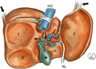

Fig. 9.2
Anatomy of the liver, inferior surface. 1 Renal and 2 colic impression on the right lobe. 3 Gastric impression on the left lobe. The star is on the gallbladder which continues in the cystic duct; the arrowhead is on the common bile duct origin; the curved arrow is on the common hepatic duct, anteriorly to which we find the common hepatic artery (red) and the portal vein (blue). The arrows are on the right and left triangular ligaments. CL caudate lobe, IVC inferior vena cava, QL quadrate lobe, RL round ligament
For its position below the diaphragm, the liver is subject to wide respiratory movements and therefore needs a valid anchorage system represented by the peritoneal reflections, otherwise referred to as ligaments.
The right portion of the coronary ligament connects the posterior face of the liver to the posterior abdominal wall; it consists of two layers, the anterosuperior and the posteroinferior ones, separated at the level of the right lobe by the bare area, and merging on the right side, into the triangular ligament. In a similar way, the anterosuperior and posteroinferior layers of the left coronary ligament merge into the left triangular ligament which connects the liver to the relevant hemidiaphragm (Fig. 9.3).
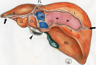

Fig. 9.3
Anatomy of the liver, posterior face. 1 Coronary ligament which continues in the right triangular ligament (arrow). 2 Coronary ligament which continues in the left triangular ligament (arrow). The liver regions uncovered by peritoneum (bare area) are highlighted with stars. The arrowhead shows the vessels of the hilum and the biliary tract enveloped in the peritoneum of the hepatoduodenal ligament. FL falciform ligament, G gallbladder, CL caudate lobe, IVC inferior vena cava and dorsal ligament, LL left lobe, inferior face, RL right lobe, inferior face
The falciform ligament, cranial portion of the coronary ligament, is a peritoneal reflection connecting the superior face of the liver to the diaphragm, from the anterior margin to the posterior abdominal wall; it looks like an almost sagittal septum, with curved borders, a convex one for the connection to the diaphragm and a concave one on the superior surface of the liver (the name derives from its shape). The falciform ligament continues caudally into the round ligament.
Finally, we find the hepatogastric and hepatoduodenal ligaments (lesser omentum), consisting of two layers of peritoneum, attaching the lesser curvature of the stomach and the duodenum to the inferomedial surface of the liver.
The liver is a special organ: it has a dual blood supply from the hepatic artery (supplying the 20–25 %) and the portal vein (supplying the 75–80 %); the refluent blood flows into the inferior vena cava through the suprahepatic veins (Fig. 9.4). The hepatic artery may originate from the celiac trunk (normally, in 65–75 % of individuals) or the superior mesenteric artery or the left gastric artery: the origin and course of such a vessel are extremely variable. The portal vein arises in the retroperitoneum from the junction of superior mesenteric and splenic veins, behind the pancreatic neck; it enters the hepatoduodenal ligament, up to the hepatic hilum; therein, the vein ramifies into two intrahepatic branches, the right and left one.
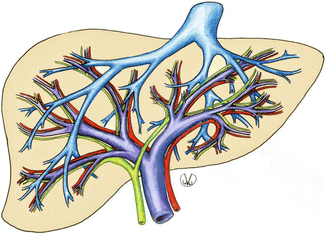

Fig. 9.4
The image shows the biliary pathways (green), the double vascular supply of the liver (red for arteries, violet for the portal system) and the drainage pathway of the hepatic veins (light blue) into the inferior vena cava
The traditional macroscopic anatomy—dividing the liver into two main lobes (right and left) and two accessory or secondary lobes (quadrate and caudate lobe) delimited by well-visible and well-defined fissures, as widely described herein above—has been replaced, in the last decades, by a more functional segmental subdivision based on the intraparenchymal distribution of the hepatic veins and portal venous and biliary branches. The portal venous, hepatic arterial, and biliary system travel together within hepatic segments and lobes (intrasegmental). The main hepatic vein courses between segments and lobe (intersegmental). Such hepatic segmental topography, obviously more complex, is also more complete and comprehensive: it provides a three-dimensional description which is the reason why it is preferred, also considering the increasing number of liver surgeries.
The middle hepatic vein divides the liver into the right and left lobe, completely independent with regard to the portal and arterial circulation and the biliary drainage. The fissure containing the middle suprahepatic vein extends from the median portion of the bed of the gallbladder, anteriorly, to the inferior vena cava, posteriorly; therefore, if in traditional anatomy the falciform ligament is the boundary between the right and left lobe, the new surgical approach considers the fissure for the gallbladder—and more precisely the Cantlie line, a straight, imaginary line going from the fissure of the gallbladder, anteriorly, to the right margin of the inferior vena cava, posteriorly—as the boundary between the two lobes. Each lobe is further divided into four segments by the right and left hepatic veins and by the right and left branches of the portal vein (the term segments has been introduced by the British authors Goldsmith and Woodburn and then spread, with subsequent amendments, by Couinaud). The current subdivision into eight segments is more effective, because each segment has an independent system of arterial supply, and venous drainage, which is essential for surgeons during hepatectomy, lobectomy, or segmentectomy.
In short, the hepatic segments are clockwise: segment 1, functionally independent, in front of the inferior vena cava, coinciding with the caudate lobe; segments 2, 3, and 4 (left lobe); and segments 5, 6, 7, and 8 (right lobe) (Figs. 9.5 and 9.6).
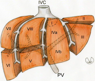
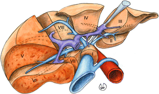

Fig. 9.5
Coronal view of the hepatic segments divided by the portal vein (PV) and the right, middle and left hepatic veins. IVC inferior vena cava

Fig. 9.6
Anatomical subdivision of the liver into hepatic segments; inferior view. The inferior vena cava is blue, the hepatic artery red, the portal vein violet
9.1.2 Biliary Tract
Bile, secreted by the hepatic cells, first flows into the bile canaliculi (microscopic ducts, contained in the portal fields of the hepatic lobule); they progressively merge together, forming interlobular biliary duct and collecting ducts that flow into the right and left hepatic ducts and then into the common bile duct. The latter, after approximately 3 cm, receives the cystic duct carrying bile to and from the gallbladder and continues into the choledochus (with a diameter of approximately 5 mm), which descends posterior and medial to the duodenum, lying in a groove on duodenal surface of pancreatic head, enters the duodenal wall, and opens up into the ampulla of Vater along with the pancreatic duct of Wirsung. A small, smooth sphincter (sphincter of Oddi) regulates the passage of bile and pancreatic juice into the duodenum.
The gallbladder, or cholecyst, is part of the biliary system; it looks like a piriform sac, on the inferior visceral surface of the liver, anterior to the right sagittal fissure. The gallbladder is 8–10 cm long, consisting of a fundus, quite dilated (about 3 cm), a conic body with a diameter of about 1–2 cm, and a neck, from where the cystic duct, approximately 3 cm long, originates. Its carrying capacity is of about 50–60 ml of concentrated bile, which flows into the duodenum when the digestion begins, thanks to the cholecystokinin stimulation (such hormone is produced by the endocrine cells of the small intestine) on the smooth muscles of the gallbladder.
9.1.3 Pancreas
The pancreas is a voluminous accessory digestive gland which passes its pancreatic fluid to the duodenum. It is located on the median line, at the level of the epigastrium and part of the left hypochondrium, deep in the abdominal cavity, at the level of the first to second lumbar vertebra, leaning on the posterior wall of the abdomen, in retroperitoneal position (anterior pararenal space); the frontal part touches the posterior surface of the stomach and the transverse colon.
Stay updated, free articles. Join our Telegram channel

Full access? Get Clinical Tree


