Fig. 11.1
Anatomy diagram of the right scrotal hemi-sac, axial section. From outside to inside, the external spermatic fascia (green), the cremaster muscle (red), the internal spermatic fascia (light blue) and the parietal and visceral laminae of the tunica vaginalis (pink) are represented. The star marks the vaginal cavity. Testis (1), spermatic cord (2)
The testis is covered by the tunica albuginea, a tough fibrous fascia that is reflected towards the posterior border of the gland to form the mediastinum testis which supports the rete testis.
From the inner surface of the tunica albuginea, a network of fibrous connective septa depart and, extending radially across the testis, reach the mediastinum and divide the parenchyma of the testis in around 250–300 lobules; these are roughly cone shaped, with their bases at the level of the tunica albuginea and the apex towards the mediastinum.
Each lobule consists of 1–4 tubuli seminiferi, each of them about 30–180 cm long; these tubes are extremely convoluted and converge towards the mediastinum where they emerge at the level of the rete testis becoming straight (tubuli recti).
The vascularization of the testes is provided by the testicular (or internally spermatic) arteries, arising directly from the aorta below the renal arteries.
At the level of the posterior margin of the testis, the veins coming from the didymus and the epididymis form convoluted larger blood vessels that anostomose in a plexus (pampiniform plexus) becoming part of the spermatic cord. The testicular vein derives from this cord and joins the inferior vena cava on the right and the ipsilateral renal vein on the left.
Lymphatic drainage of the testes follows the testicular arteries back to the pre- and para-aortic lymph nodes.
11.1.2 Pathway of Sperm
The pathway of sperm begins in the testis with the tubuli recti and the rete testis and continues with the epididymis, the ductus deferens, the ejaculatory duct and, finally, the urethra.
Around 10–15 small calibre efferent ducts originate from the rete testis and emerge from the posterosuperior surface of the testis to form the head of the epidydimis. This organ lays in a posterosuperior position in respect to its ipsilateral didymus and has the function to store the spermatozoa and facilitate their maturation. Each epididymis has the shape of a large comma and can be divided in three main regions: the head, the enlarged superior end; the body, the intermediate portion; and the tail, the lower end that continues with the vas deferens (Fig. 11.2).
Fig. 11.2
Anatomic diagram of the testis (1), the epididymis (2), the ductus deferens (3) and the pampiniform plexus (4). The sagittal section shows the tunica albuginea (arrows) and the septa (arrowheads), between which lie the seminiferous tubules which converge towards the mediastinum testis. At this level the efferent ductules (curved arrow) draining into the head of the epididymis (star) can be recognized
A very convoluted conduit (duct of epididymis) originates from the union of the efferent ducts in the head of the epididymis.
The tail of the epididymis continues with the vas deferens (or ductus deferens) that has a roughly cylindrical shape, a 2–3 mm calibre and a length of about 40 cm. It is divided in several parts (testicular, funicular, inguinal and pelvic). The testicular part of the vas deferens runs in proximity of the posterior face of the tail and the body of the epididymis; at the limit between the head and the body of the epididymis, the ductus deferens distances itself from this organ and continues upwards to become part of the spermatic cord.
Besides the vas deferens, which is located in the posterior part, the spermatic cord contains the testicular, deferential and cremasteric (or external spermatic) arteries; testicular veins (pampiniform plexus); lymphatic vessels; nerves; the vaginal ligament and the internal cremaster muscle.
Within the spermatic cord, the ductus deferens (inguinal part) runs through the inguinal canal. When it reaches the internal abdominal orifice, the ductus deferens leaves from the other elements of the spermatic cord and running through the extraperitoneal pelvic space (abdomino-pelvic part) reaches the posterior part of the urinary bladder, passes over and medially the ureter and continues medially descending towards the base of the prostate where it converges with the contralateral ductus. At this level, the union of the ductus deferens with the duct of the ipsilateral vesicle gland forms the ejaculatory duct.
The ejaculatory ducts are about 2–2.5 cm long and run almost entirely within the prostate, emerging through two small orifices on the posterior wall of the prostatic urethra, where a longitudinal elevation called seminal colliculus or verumontanum is located.
The male urethra is 18–20 cm long; in the initial tract, it constitutes a passage for urine only, whereas in the part going from the orifices of the ejaculatory ducts to the external urethral meatus, it also transports sperm. The urethra is divided in three parts: prostatic, about 3 cm long; membranous, about 1.5 cm long, located deeply within the urogenital diaphragm; and spongy or cavernous, the longest part (13–15 cm), sheathed by the urethral corpus spongiosum. The spongy urethra is subdivided in bulbous, penile and navicular and opens onto the external urethral meatus; the ducts of the urethral glands (glands of Littre) emerge in its wall (Fig. 11.3).
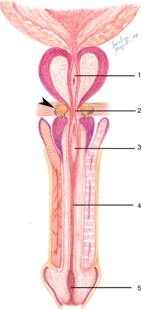
Fig. 11.3
Anatomy of the male urethra. Coronal section. The different portions of the urethra can be visualized: prostatic (1), membranous (2), bulbar (3), penile (4) and navicular portion (5). The arrowhead indicates the Cowper glands
Within the urogenital diaphragm there are also the bulbourethral glands (also Cowper’s glands) and their ducts enter the urethra in the initial tract of the spongy part.
11.1.3 Seminal Vesicles
The seminal vesicles are a pair of small symmetrical glands that are poorly enveloped in children and atrophic in elderly men; in adults, their main axis, oriented downwards, forward and medially, measures 5–8 cm. The vesicles are positioned in the pelvis between the base of the urinary bladder and the rectum and are related to the prostate posterosuperiorly; in their prevesical tract, ureters runs medially to the vesicles. Each seminal vesicle unites with its respective ipsilateral vas deferens at an acute angle to form the ejaculatory ducts (Fig. 11.4).
Fig. 11.4
Anatomy of the distal vas deferens, the seminal vesicles and the prostate. The posterior view visualizes the anatomical relationships among the ductus deferens and the ampulla of ductus deferens (1), the prostate (2), the ureter (3), the urinary bladder (4) and the seminal vesicles (5)
11.1.4 Prostate
The prostate is a glandular organ located medially in the lesser pelvis (extraperitoneal pelvic space) between the base of the urinary bladder and the urogenital diaphragm, behind the pubic symphysis and in front of the rectal ampulla, between the medial bundles of the two elevator muscles of the anus (pubococcygeus muscles); the prostate is shaped like a chestnut, with an apex pointing downwards and forward and a base turned upwards and backwards (Fig. 11.5).
Fig. 11.5
Anatomy of the male genital system, sagittal view. The anatomical relationships of the prostate (1) with the urinary bladder (2), the seminal vesicles (3) and the rectum (4) are shown. Corpora cavernosa (5), corpus spongiosum (6), glans (7)
The prostate has a hard-elastic consistency. It is very small in the child and starts growing only during puberty reaching, around 20–25 years of age, a height of 3 cm, a width of 4 cm (at the base) and a thickness of 2.5 cm. At this age its weight is around 20 g.
The prostate is surrounded by a layer of muscular fibres and collagen that represents a capsule. At the front, it is connected to the pubic symphysis by two bands of connective tissue named puboprostatic ligaments; behind, it is separated from the rectum by the Denonvilliers’ fascia; in the lower part, the apex of the prostate borders on the genitourinary membrane.
Crossing through the prostate, from above to below, is the first portion of the urethra (prostatic urethra) into which the prostate empties its secretions during ejaculation. A sharp angle of 145°, between the base and the apex of the prostate, divides the urethra in two segments, proximal and distal, having nearly the same length but completely different anatomical relationships. A first sphincter formed by a cylinder of smooth muscular fibres surrounds the proximal segment of the urethra between the neck of the urinary bladder and the base of the verumontanum; the only function of this preprostatic sphincter is probably preventing the retrograde flux of semen by contracting during the ejaculation. The verumontanum is located inthe distal segment. This part is covered by a second semicylindrical sphincter formed by striated muscular fibres that continues distally with the external striated sphincter, beyond the apex of the gland. In the posterosuperior part, the prostate is also crossed, obliquely, by the ejaculatory ducts.
The gland can be subdivided in four lobes according to the different relationships with the urethra and the ejaculatory ducts established by the various parts: anterior, medium and two lateral lobes. The anterior lobe lies in front of the urethra, the medium lobe is located between the urethra and the ejaculatory ducts and the two lateral lobes (left and right), most voluminous, continue behind the frontal plan passing through the ejaculatory ducts.
The anatomical subdivision proposed by McNeal considers the heterogeneous composition of the prostate that includes also purely muscular or fibromuscular regions, besides glandular ones. Each region drains a specific urethral segment and shows some histological differences from the others.
According to McNeal’s description, the urethra separates roughly the gland in an anterior (ventral fibromuscular) and posterior (dorsal glandular) portion.
The ventral fibromuscular stroma is mainly composed by smooth muscular fibres that are a continuous with the fibres of the detrusor muscle of the anterior wall of the urinary bladder. In fact, these fibres extend distally from the neck of the urinary bladder and expand laterally covering the whole anterior and anterolateral surface of the glandular prostate. The thickness of the muscular tissue increases distally until the verumontanum, where its mass is further incremented by a fibrotic component. Caudally to the verumontanum, the muscular tissue becomes progressively thinner towards the prostatic apex.
The dorsal glandular portion is subdivided into external and internal region as well. The first region includes the central and the peripheral zones which account for approximately the 25 % and the 75 % of the volume of the prostate, respectively, and are markedly distinct from each other both macro- and microscopically, in particular in the normal gland in young individuals. The central zone surrounds the posterosuperior part of the proximal urethra and the entire course of the ejaculatory ducts so that the orifices of the latter are encircled by those of the central zone on the rounded apex of the verumontanum. The peripheral zone surrounds both the central zone and the distal tract of the urethra; its principal ducts open in a double lateral line in the distal segment of the urethra, separately from those of the central zone.
The internal region includes the transitional (around 5 % of the volume of the gland in young individuals) and the periurethral (less than 1 % of the glandular prostate) zones. The transitional zone is represented by two small lobules located just laterally to the preprostatic sphincter. Its ductal orifices open along the proximal continuation of the double lateral line formed by the orifices of the ducts of the peripheral zone. The glands of the periurethral zone are confined in the stroma immediately adjacent to the proximal urethra; their ductal orifices open in the proximal urethral segment in a double lateral line and represent the further proximal continuation of the ductal systems of the peripheral and transitional zones (Figs. 11.6 and 11.7).
Fig. 11.6
Diagram of the zonal anatomy of the prostate. The coronal section shows the peripheral (light blue), central (brown) and transitional (violet) zones. Urethra (1). The arrowheads indicate the periurethral glands
Fig. 11.7


Schematic drawing of the zonal anatomy of the prostate. The sagittal section shows the peripheral (light blue), central (brown), transitional (violet) and anterior fibromuscular (yellow) zones. Urethra (1). The arrowhead indicates the ejaculatory duct
Related posts:

Stay updated, free articles. Join our Telegram channel
Full access? Get Clinical Tree
