35.1 CT Abdomen: upper abdomen
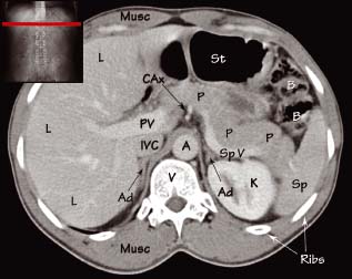
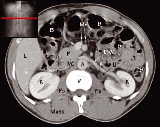
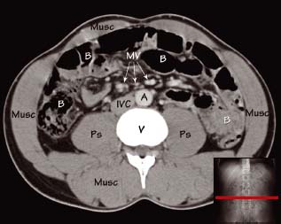
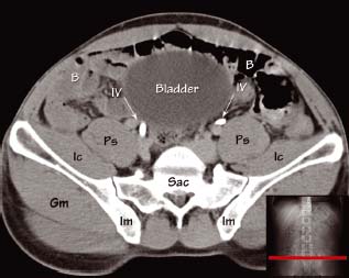
Key
A – Aorta
Ad – Adrenal gland
B – Bowel
CAx – Coeliac axis/trunk
Gm – Gluteus muscles
Ic – Iliacus muscle
Im – Ilium
Iv – Iliac vessels
IVC – Inferior vena cava
K – Kidney
L – Liver
Musc – Muscles of abdomen and back
MV – Mesenteric vessels
P – Pancreas
Ps – Psoas muscle
PV – Portal vein
Sac – Sacrum
Sp – Spleen
SpV – Splenic vein
St – Stomach
U – Ureter
V – Vertebral body
Abdominal anatomy seen on CT
CT is superior to plain X-ray imaging in demonstrating abdominal anatomy. Data is usually acquired during the portal venous phase of contrast agent enhancement (approximately 60 – 70 seconds post-injection) to optimise delineation of the liver, which derives its primary supply from the portal vein. Important structures visible on abdominal CT include:
- Stomach – this left upper quadrant hollow structure extends across the epigastrium. It is divided into the fundus, body, antrum and pylorus, and its blood supply is derived from the coeliac trunk.
- Small bowel –
Related posts:

Stay updated, free articles. Join our Telegram channel


Full access? Get Clinical Tree


