36.1 Colorectal cancer (CT with rectal contrast)
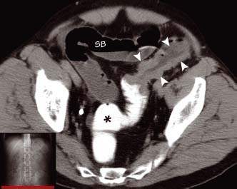
A segment of bowel wall shows circumferential irregular thickening (arrowheads). Rectal contrast material (*) introduced via a rectal catheter could not pass beyond the lesion which has obstructed the bowel. Proximal to the lesion there is dilatation of both large and small bowel (SB)
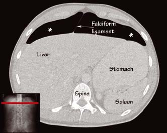
Lung window settings are highly sensitive for the presence of free intra-abdominal gas. This patient suffered an injury to the bowel from a high-speed road accident. Air (*) is seen either side of the falciform ligament separating the liver from the anterior abdominal wall
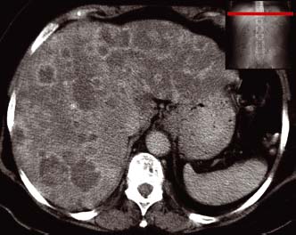
Numerous low-density irregular nodules are seen in the liver. Many of these show contrast enhancement around their rim. This patient with metastatic breast cancer had a recent ultrasound examination which had underestimated the volume of disease
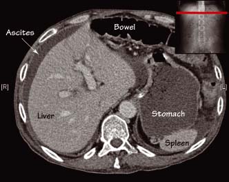
A crescent of low-density material is seen around the edge of the liver, which has a similar density to the contents of the stomach. This patient has free intra-abdominal fluid (ascites) due to peritoneal metastases from an ovarian carcinoma
Bowel pathology
- Inflammatory bowel disease (IBD) (see Chapter 18) – CT is useful in evaluating IBD. In Crohn’s disease CT can show thickened bowel wall and intra-abdominal complications (e.g. fistulae, abscesses, small bowel obstruction). Due to the predilection for Crohn’s disease to affect the small bowel, CT enteroclysis is being increasingly used to depict Crohn’s disease (i.e. a volume challenge of an enteral contrast agent is administered via a nasojejunal catheter before CT imaging to distend the small bowel and improve the detection of mural abnormalities). The CT findings in ulcerative colitis are primarily thickening of the large bowel wall, which begins distally and spreads proximally in a continuous fashion.
Stay updated, free articles. Join our Telegram channel

Full access? Get Clinical Tree


