37.1 Pancreatic cancer
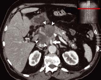
A lobulated low-density mass is seen in the head of the pancreas (arrowheads). Normal contrast enhancement of the body and tail of the pancreas is demonstrated (*). The mass has engulfed the superior mesenteric vessels (arrow) making it unresectable
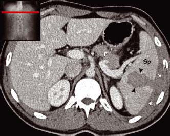
This patient had been in a road traffic accident and sustained blunt injury to the left side of the abdomen and chest. Here there is a large splenic laceration (arrowheads) with a contained splenic capsular haematoma (*). The patient was haemodynamically stable and no active extravasation of contrast is seen. Sp spleen, P pancreas
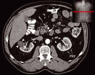
A small irregular low-density soft tissue mass (arrowheads) is arising from the lateral edge of the right kidney (RK). Despite its small size there has been breach of the perirena fascia and the mass approaches the liver. Compare this with the left kidney (LK) and the left-sided well-defined perirena fascia (PRF)
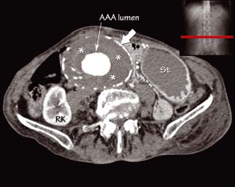
There is a very large abdominal aortic aneurysm (AAA). The outer wall is calcified (arrow) and within this there is a thick wall of soft tissue density thrombus (*). Contrast is seen flowing through the central lumen. St stomach, RK right kidney
Pancreatic lesions
- Acute pancreatitis
Related posts:


Stay updated, free articles. Join our Telegram channel

Full access? Get Clinical Tree


