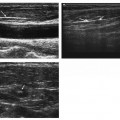
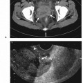
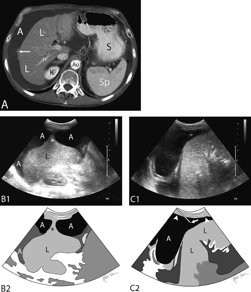
16 Abdominal Paracentesis Fig. 16.1 Paracentesis performed from adjacent to the liver that has an adhesive band between the right liver lobe and the peritoneum. (A) Axial computed tomography (CT) image with contrast enhancement at the level of the aortic hiatus. There appears to be tenting of the hepatic parenchyma toward the peritoneum at the midaxillary line (arrow). Occasionally the majority, if not all, of the ascites fluid is perihepatic, which requires a right upper quadrant access paracentesis rather than the more common lower quadrant sites. It is important to avoid the liver capsule and/or mistaking a large and distended gallbladder for a loculated fluid collection (A, ascites; L, liver; IVC, inferior vena cava; H, right hepatic vein; Ao, aorta; S, stomach; Sp, spleen). (B) Gray-scale ultrasound image of the upper abdomen (top) and schematic sketch of it (bottom). The band is tenting toward the peritoneum (*). The operator avoided the band. In addition, the operator did not go deep with the needle avoiding transgression of the liver capsule (A, ascites; L, liver). (C) Gray-scale ultrasound image of the upper abdomen (top) and schematic sketch of it (bottom). The 21-gauge access needle (arrow) is seen traversing the peritoneum (arrowheads). The operator avoided the band. In addition, the operator did not go deep with the needle avoiding transgression of the liver capsule (A, ascites; L, liver).
Indications
Contraindications
Relative Contraindication
Preprocedural Evaluation
Evaluate Prior Cross-Sectional Imaging
Evaluate Preprocedure Laboratory Values
Obtain Informed Consent
Equipment
Ultrasound Guidance
Standard Surgical Preparation and Draping
Local Infiltrative Analgesia Administration
Sharp Access Devices
Tubular Access Devices
Stay updated, free articles. Join our Telegram channel

Full access? Get Clinical Tree


