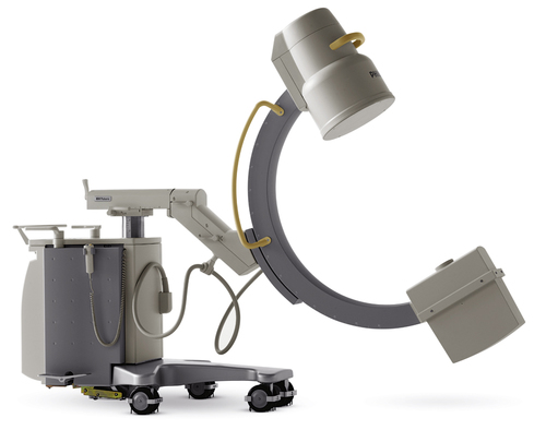
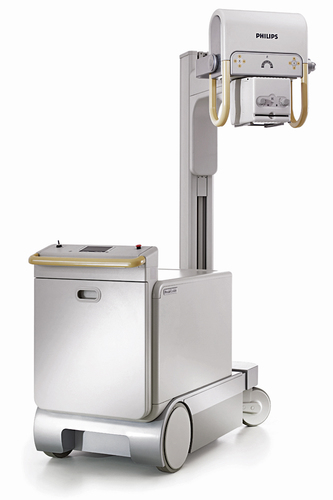
• Differentiate between radiographic and fluoroscopic mobile equipment. • State the purpose of dedicated units and identify their unique features. • Explain the principles of linear tomography. • Recognize the variations required in exposure technique factors for mobile and dedicated units, and linear tomography. Mobile x-ray units can be smaller light-duty units or full-power units that are transported on wheels (Figure 15-1). These mobile units may be transported with or without the use of motors. Mobile units can be categorized in several ways depending on the design of the generator: direct power, battery power, capacitor-discharge, or high-frequency. Mobile units operated by plugging into a wall outlet for direct power may experience fluctuations in voltage, which affects the radiation output. Battery-operated units provide more consistent radiation output, similar to a three-phase generator, but need to be recharged. Capacitor discharge units must be plugged into a wall outlet during operation, but produce consistent radiation output, similar to a single-phase generator. High-frequency units produce a consistent radiation output and are lightweight, but must be plugged into a wall outlet during operation. C-arm mobile units have fluoroscopic capabilities that are typically used in the operating room when imaging is necessary during surgical procedures. A video unit is also attached, which offers both static and dynamic recording during the procedure. Because it is a fluoroscopic system, many of the features of a fixed fluoroscopic unit are also made available with a C-arm. A C-arm unit is designed with an x-ray tube and image intensifier attached in a C configuration (Figure 15-2). As a result, the unit can be positioned in a variety of planes, enabling viewing from different perspectives. Generally, three sets of locks are provided to move and hold the C-arm in place. One set moves the entire “c” toward or away from the base (the equivalent of moving a table side to side). Another set allows the pivot of the “c” about its axis (the equivalent of angling a general radiographic tube head assembly). The last set allows the “c” to slide along its arc (the equivalent of moving the patient from anteroposterior or posteroanterior to oblique to lateral without having to move the patient).
Additional Equipment
Mobile Equipment
Radiographic Units
Fluoroscopic Units
![]()
Stay updated, free articles. Join our Telegram channel

Full access? Get Clinical Tree


Additional Equipment
 Critical Concept 15-1