Adrenal Glands, Pancreas, and Other Retroperitoneal Structures


Adrenal Glands, Pancreas, and Other Retroperitoneal Structures


The retroperitoneum is bordered superiorly by the diaphragm, inferiorly by the pelvic brim, anteriorly by the parietal peritoneum, and posteriorly by the transversalis fascia. Located within this compartment are larger structures, such as the adrenal glands, pancreas, kidneys and ureters, duodenal loop and ascending and descending colon, and the great vessels and their branches, as well as smaller structures, such as the lymph nodes, lymphatic channels, and nerves. This chapter will review the normal anatomy and common pathologic disorders of the adrenal glands, pancreas, lymph nodes, major vascular structures, and soft tissues. The psoas muscles, which actually lie within the retrofascial space posterior to the retroperitoneum, are included in the discussion because conditions affecting these structures often involve the retroperitoneal compartment. Diseases of the kidney and duodenum and colon have been covered elsewhere in this book (Chapter 7).


ADRENAL GLANDS


NORMAL ANATOMY

With high-quality techniques, both limbs of the adrenal glands can be seen on sonography, computed tomography (CT), and magnetic resonance imaging (MRI) (1,2,3). Characteristically, the adrenal glands have an inverted Y or V shape (Fig. 9.1). Occasionally, on CT the right adrenal gland may appear as a linear structure next to the diaphragmatic crus. This appearance is the result of visualization of only the medial limb of gland. The lateral limb may not be seen if it is closely apposed to the liver.


In neonate and infants, the adrenal glands are relatively large structures, up to one-third the size of the kidneys, due to a relatively larger volume of cortex (1). There is no
significant difference in the size of the two glands. The limbs of the adrenal glands have a uniform thickness, except at the apex of the gland where they unite. The surface of the adrenal glands should be smooth, without nodular protuberances, and the thickness of the limbs should be uniform.


Figure 9.1 Neonatal adrenal glands, normal sonographic shapes. A. Transverse sonogram of left adrenal gland shows an inverted V configuration. B. Transverse sonogram showing the inverted Y configuration. The medulla is echogenic and is surrounded by hypoechoic cortex. C. Arterial phase contrast-enhanced CT in another infant shows both limbs of the right adrenal gland (arrowheads) with enhancement of the cortex. A nasogastric tube (arrows) is present in the stomach.

The sonographic appearance of the normal adrenal gland varies with age. In newborns, the adrenal medulla is relatively thin and hyperechoic and is surrounded by thicker hypoechoic cortex (see Fig. 9.1). The adult appearance is acquired between 1 and 3 years of age. At this stage, the adrenal glands are seen as very thin, linear, hypoechoic structures. On CT, the adrenal glands have soft-tissue attenuation similar to that of the liver. On spin-echo T1-weighted MR images, the adrenal glands have intermediate signal intensity similar to that of the liver and less than that of fat. On T2-weighted and fat-suppressed images, the adrenal glands are slightly brighter than liver and much brighter than suppressed fat. The normal adrenal glands may demonstrate transient enhancement after intravenous administration of contrast medium or gadolinium.


CONGENITAL ANOMALIES

Congenital absence of the adrenal glands is extremely unusual. Virtually all patients with renal agenesis or ectopia have an ipsilateral adrenal gland, but the gland elongates and assumes a flattened or discoid configuration parallel to the spine (Fig. 9.2). By comparison, the adrenal glands have a normal appearance in patients who have had a nephrectomy.


Figure 9.2 Adrenal gland in a newborn with renal agenesis. Longitudinal sonogram shows an elongated and discoid right adrenal gland (arrowheads). Corticomedullary differentiation is preserved.

Two anomalies of adrenal fusion have been described. In the circumrenal gland, the fused limbs surround the upper pole of the kidney. In the horseshoe gland, the two limbs of the gland are fused in the midline anterior to the spine and are connected by an isthmus of tissue (Fig. 9.3).


NEOPLASMS

Sonography is performed to confirm the presence of a clinically suspected mass and its site of origin. After the presence of an abdominal mass has been confirmed by sonography, patients undergo further imaging with either CT or MRI. Positron emission tomography (PET) and metaiodobenzylguanidine (MIBG) scintigraphy are also used in the staging of neuroblastoma.


Figure 9.3 Fusion anomalies. A. Circumrenal adrenal gland. Transverse sonogram shows the adrenal gland (arrowheads) encircling the upper pole of the left kidney (K). B. Horseshoe adrenal. Transverse scan shows the isthmus (arrow) connecting the right and left adrenal glands (arrowheads) (A, aorta; S, spine).


Neuroblastoma

Neuroblastoma is the most common solid, extracranial malignant tumor in children, accounting for 8% to 10% of all childhood cancers (4,5,6). Most children with neuroblastoma are between 1 and 5 years of age, with a median age at diagnosis of 22 months. Neuroblastomas may arise in the adrenal medulla or anywhere along the sympathetic ganglion chain. Up to 75% of neuroblastomas arise in the abdomen, and two-thirds of these lesions arise in the adrenal medulla. The remaining abdominal and pelvic tumors usually originate in the paravertebral sympathetic ganglia or presacral area. Occasionally, an abdominal tumor arises in the organ of Zuckerkandl. On cut-section, neuroblastoma is usually well demarcated, although it lacks a capsule, and it commonly contains areas of necrosis, hemorrhage, cystic degeneration, and calcification. Microscopically, the tumor is composed of small, darkly straining, round cells that are characteristically arranged in rosettes. Average tumor size is 8 cm at the time of presentation.


The most common clinical presentation of abdominal neuroblastomas is an abdominal mass. Other presenting features include skeletal pain; neurologic abnormalities, such as nystagmus, ataxia, opsoclonus, and paraparesis; ophthalmic signs, including proptosis and periorbital ecchymoses; and gastrointestinal complaints, such as diarrhea, anorexia, and vomiting. These signs and symptoms usually are related to metastatic spread of the tumor, intraspinal invasion, or the production of various hormones. Approximately 90% of patients with neuroblastoma have increased serum or urinary levels of catecholamines or their metabolites, particularly vanillylmandelic acid (VMA) and homovanillic acid (HVA). Between 60% and 70% of children have metastatic disease at time of diagnosis. Common sites of metastases are distant lymph nodes, cortical bone, bone marrow, liver, and skin. Neuroblastoma has been associated with neurofibromatosis type I and with aganglionosis of the colon.


Two paraneoplastic syndromes have been described in association with neuroblastoma. One is myoclonic encephalopathy of infancy, which includes the triad of cerebellar ataxia, myoclonus, and opsomyoclonus. The precise cause of this syndrome is unknown, but it has been postulated that an immune response to the primary tumor leads to production of antineural antibodies that cross-react with cerebellar tissue. The second syndrome consists of intractable watery diarrhea and hypokalemia caused by excess production of vasoactive intestinal peptides.


Table 9.1: International Neuroblastoma Staging System


Stage I


Localized tumor confined to the area of origin; complete gross excision, with or without microscopic residual.


Stage IIA


Unilateral tumor with incomplete gross excision; ipsilateral and contralateral lymph nodes negative microscopically.


Stage IIB


Unilateral tumor with complete or incomplete gross excision with positive ipsilateral regional lymph nodes; identifiable contralateral lymph nodes negative microscopically.


Stage III


Tumor infiltrating across the midline with or without regional lymph node involvement; or unilateral tumor with contralateral regional lymph node involvement; or midline tumor with bilateral regional lymph node involvement.


Stage IV


Dissemination of tumor to distant lymph nodes, bone marrow, liver and/or other organs (except as defined in stage IVS).


Stage IVS


Localized primary tumor as defined for stage I or II with dissemination limited to liver, skin, and/or bone.


The International Neuroblastoma Staging System (INSS), based on the combination of clinical, radiographic, and surgical findings, is the system used most often to stage neuroblastoma (see Table 9.1). Distribution of stages based the INSS is approximately I, 20%; II, 10%; III, 15%; IV, 50%; and IV-S, 4% (5).

The treatment of neuroblastoma varies with the stage of the tumor. Patients with tumors that are localized to one side of the midline or cross the midline without encasement of major vessels undergo primary surgical resection. Chemotherapy is the treatment of choice in patients with unresectable disease. The use of radiation therapy is limited to patients with localized disease that cannot be resected and does not regress completely with chemotherapy. Once unresectable tumors decrease sufficiently in volume, delayed surgical resection is performed. Infants with IV-S disease may not be treated, as spontaneous regression sometimes occurs in these patients.

The principal determinants of prognosis are the stage and site of the tumor and the age of the patient. Tumors with lower stages, those arising at extra-abdominal sites, and tumors occurring in children under 1 year of age have a more favorable prognosis. Stage is a particularly important factor. Two-year survival ranges from 80% with disease limited to the adrenal to less than 5% for disease with skeletal metastases. Thoracic tumors also have a better overall survival rate than abdominal tumors. Several biological markers of tumor tissue also influence survival rate. Favorable factors include triploid karyotypes, well-differentiated stroma, an unamplified N-myc oncogene, and absence of abnormalities of chromosome 1. Unfavorable prognostic signs are a “diploid” (decreased DNA) karyotype, N-myc amplification (more than 10 copy numbers), and allelic loss of chromosome 1p.


Imaging Evaluation


Plain Radiography.

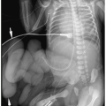
Plain films may be acquired for unrelated clinical indications and demonstrate an unsuspected primary tumor in the chest or abdomen or they may be obtained to further evaluate a suspected or known abdominal mass (most often diagnosed by sonography). Plain radiographic findings of abdominal neuroblastoma include a paraspinal mass and enlargement of the intervertebral foramina or erosion of the pedicles due to intraspinal extension of tumor. The tumor may contain calcifications.


Sonography.

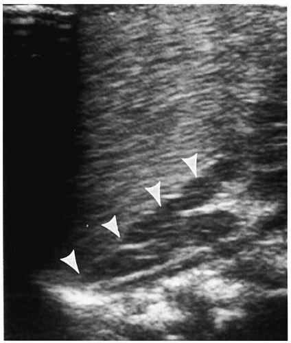
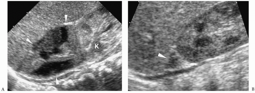
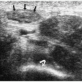
The evaluation of patients with palpable abdominal masses, including neuroblastoma, usually begins with sonography (1,6,
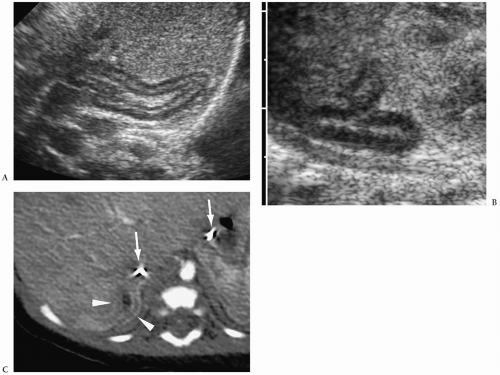
7). Neuroblastoma appears as a suprarenal or paraspinal mass. It may be homogeneous or heterogeneous and it may contain hyperechoic areas representing calcification and hypoechoic areas as a result of hemorrhage, necrosis, or cystic degeneration (Fig. 9.4). Tumor margins may be smooth or irregular. Peripheral or central vascularity may be observed on color Doppler sonography.


Figure 9.4 Neuroblastoma. A. Longitudinal sonogram shows a heterogenous suprarenal mass (arrowheads) just inferior to the stomach (ST). The mass contains scattered hyperechoic areas representing calcification. (Renal, left kidney). B. Longitudinal sonogram in another patient shows a heterogeneous mass (cursors) adjacent to the aorta (A). The tumor arose in the sympathetic ganglia.


Figure 9.5 Newborn neuroblastoma. Contrast-enhanced CT scan shows a cystic right suprarenal mass (M), which has displaced the kidney inferiorly.

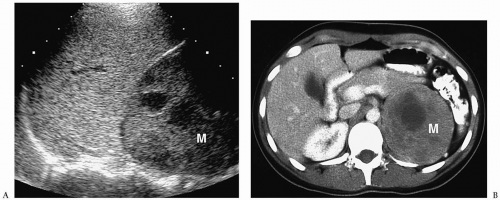
In the newborn, neuroblastomas may be predominantly cystic or anechoic (8,9) (Fig. 9.5). On pathologic sections, this appearance reflects either degenerative change in the tumor or, in some cases, clusters of microcysts in the tumor cells.


Computed Tomography.

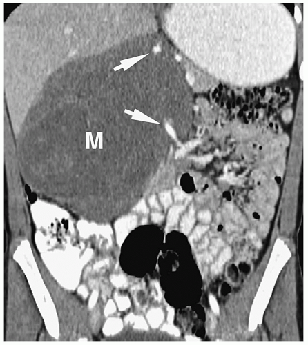
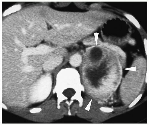
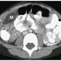
On CT, neuroblastoma appears as a homogeneous or heterogeneous, soft tissue mass with irregular margins (2,4,6,7). The tumor enhances less than that of surrounding tissues after intravenous administration of contrast material. Calcifications occur in approximately 85% of abdominal neuroblastomas (Fig. 9.6).


Magnetic Resonance Imaging.

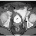
Neuroblastoma has low or intermediate signal intensity on T1-weighted sequences and high signal intensity on T2-weighted and fat-suppressed sequences (4,6,10,11). It usually enhances after administration of intravenous gadolinium chelate compounds (Fig. 9.7). Calcification has low signal intensity on all imaging sequences. Areas of necrosis and cyst formation are usually hypointense on T1-weighted images and hyperintense on T2-weighted sequences and do not enhance following gadolinium administration.


Figure 9.6 Neuroblastoma. Coronal reformatted CT scan in a 2-year-old boy shows a large mass (M) arising from the right adrenal, crossing the midline and encasing the superior mesenteric artery (arrows).


Figure 9.7 Adrenal neuroblastoma. A. T1-weighted coronal image demonstrates a low signal intensity mass (M) arising from the left adrenal gland, displacing the celiac axis (arrow) (K, kidney). B. On the postgadolinium image, the mass (M) enhances heterogeneously. C. A fat-saturated fast spin-echo T2-weighted image from another patient shows a right adrenal mass (M) extending posterior to the inferior vena cava (C), crossing the midline, and surrounding the celiac axis (arrowheads) (LK, left kidney).


Imaging Intra-abdominal Tumor Spread.

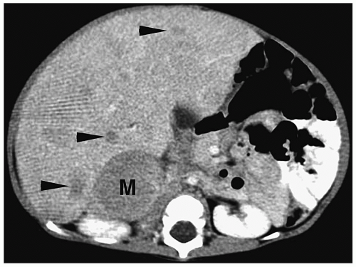
Patterns of intra-abdominal extension of tumors include midline extension, vessel encasement, spread to regional lymph nodes, intraspinal extension, and hepatic metastases. Inferior vena caval and aortic displacement are common. Bone metastases also may be seen.

Hepatic metastases have a variable appearance. The lesions can be discrete and solitary or multifocal. Discrete hepatic lesions are hyperechoic or hypoechoic on sonography, low attenuation on CT, hypointense to adjacent liver on T1-weighted MR images, and hyperintense on T2-weighted and fat-suppressed images. Diffuse metastatic infiltration produces hepatomegaly with heterogeneity of the hepatic parenchyma (Fig. 9.8).


Imaging Distant Tumor Spread.

Distant spread is most commonly to lymph nodes and skeleton. A common site of distant nodal spread is the supraclavicular region.


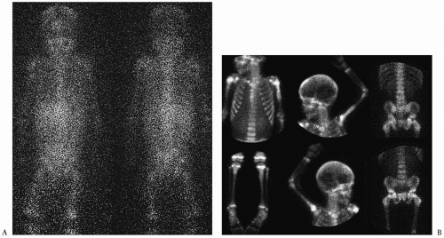
Skeletal metastases occur in 50% to 60% of patients at diagnosis, mostly in patients older than 1 year of age (5). These can involve cortical bone or bone marrow. MIBG has become the study of choice to image skeletal metastases (12) (Fig. 9.9). Abnormal activity can be seen in the primary tumor and in bone, bone marrow and soft tissue metastases. PET performed with the glucose analogue 2-[fluorine-18]-fluoro-2-deoxy-D-glucose (FDG) has been used most recently for the evaluation of distant disease extent. Most neuroblastomas and their metastases avidly concentrate FDG prior to chemotherapy or radiation therapy (13). Skeletal metastases also can be seen on MRI, where they have a low signal intensity on T1-weighted sequences and high signal intensity on T2-weighted and fat-suppressed sequences.


Figure 9.8 Hepatic metastases in an infant with stage IVS neuroblastoma. CT scan shows multiple low attenuation lesions (arrowheads) and a soft tissue mass (M) in the right adrenal gland.


Follow-up Imaging.

CT, MRI, MIBG, and PET have been used to assess the response of the tumor to surgery, chemotherapy, and radiation therapy. Neuroblastoma tends to regress in size with treatment, although the regression may be incomplete, leaving a residual retroperitoneal soft-tissue mass.


Ganglioneuroblastoma and Ganglioneuroma

Ganglioneuroblastoma is a malignant tumor containing both primitive and differentiated cells. Ganglioneuroma is a benign completely differentiated tumor composed of mature ganglion cells. Ganglioneuroblastoma and ganglioneuroma tend to occur later in the first decade of life or in the second decade of life. They have imaging findings similar to those of
neuroblastoma (6). The diagnosis is made histologically by the degree of cellular maturation and differentiation.


Figure 9.9 Metastatic neuroblastoma. A. Anterior scintigram obtained 48 hours after injection of 123I-MIBG demonstrates increased activity in the skeleton consistent with bone and bone marrow metastases. B. Bone scintigraphy at the same time as MIBG scan demonstrates widespread osseous metastases most prominent in the ends of long bones, spine, and skull.


Pheochromocytoma

Pheochromocytoma is a functionally active neoplasm of the adrenal medulla that causes catecholamine overproduction. Most pheochromocytomas originate in the adrenal medulla, but up to 30% may arise in extraadrenal sites, mainly the paravertebral sympathetic chain, paraaortic bodies, and bladder wall. When this tumor originates outside of the adrenal glands, it is termed a paraganglioma. Most pheochromocytomas in children are benign.


Clinical features suggestive of the diagnosis are paroxysmal hypertensive episodes, headache, tachycardia, palpitation, diaphoresis, and pallor. Patients with bladder wall paragangliomas may present with micturition syncope. There is an increased incidence of pheochromocytomas in patients with multiple endocrine neoplasia type 2 (medullary thyroid carcinoma and parathyroid disease), and in patients with neurofibromatosis and von Hippel-Lindau disease. Multiple or bilateral tumors are more likely in these syndromes. The diagnosis of pheochromocytoma is usually established by biochemical testing, which demonstrates elevated urinary or serum catecholamines levels or elevated urine metanephrine or vanillylmandelic acid levels.

Localization of pheochromocytomas is accomplished with CT, MRI, or 123I- or 131I-labeled MIBG. Total body scanning with MIBG is especially useful in patients with multifocal tumors or metastatic disease.

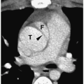
Pheochromocytomas are usually large (>2 cm in diameter), sharply marginated lesions (1,2,3,4,11) (Fig. 9.10). Smaller tumors commonly have a homogeneous matrix, whereas larger tumors are often heterogeneous. Calcifications are present in about 15% of tumors. The tumors are predominantly soft-tissue attenuation on CT, hypointense to liver on T1-weighted images, and markedly hyperintense to liver and fat on T2-weighted and fat-suppressed images. Moderate to marked heterogenous enhancement after intravenous administration of contrast medium or gadolinium is typical. Signs of malignancy, such as local invasion, lymph node enlargement and distant metastases, may also be seen. The imaging appearance of pheochromocytoma is not specific and differentiation from other adrenal tumors requires biochemical studies.


ADRENOCORTICAL TUMORS

Adrenocortical lesions in children are usually hyperfunctioning. Less commonly, they are nonfunctioning and present as palpable abdominal masses (14,15,16).


Figure 9.10 Pheochromocytoma. Contrast-enhanced CT shows a 3-cm heterogeneous left adrenal mass (arrowheads) with central necrosis. The soft tissue components show moderate enhancement. (From Siegel MJ, ed. Pediatric body CT. Philadelphia. Lippincott Williams & Wilkins 1999).


Figure 9.11 Adrenal carcinoma. A 12-year-old girl with hirsutism. A. Longitudinal sonogram shows a heterogeneous left suprarenal mass (M). B. Contrast-enhanced CT reveals a large mass (M) with areas of low attenuation as a result of necrosis.


Adrenocortical Cancer

Adrenal carcinoma is a rare highly malignant neoplasm of the adrenal cortex. The tumor commonly causes virilism in girls and pseudoprecocious puberty in boys. Cushing syndrome, feminization, and hyperaldosteronism are less common presentations. Mean patient age at diagnosis is 9 years. The tumor may invade the inferior vena cava and spread to regional lymph nodes. Distant metastases are to lung, liver, and lymph nodes. Biochemical criteria for diagnosis of a virilizing tumor are elevated levels of urinary 17-ketosteroids and normal to mildly elevated urinary cortisol levels.


Adrenal carcinomas are readily detected by sonography, CT, and MRI. They have an imaging appearance similar to that of neuroblastoma. Most often they are seen as large masses, measuring more than 5 cm in diameter, at the time of presentation. Central areas of necrosis are common. Calcification is found in 25% to 40% of tumors (Fig. 9.11). They are echogenic on sonography, soft-tissue attenuation on CT, hypointense to liver on T1-weighted images, and hyperintense to liver on T2-weighted and fat-suppressed images. Adrenal carcinoma enhances after administration of intravenous contrast medium and gadolinium.


Adrenal Adenomas

Adrenal adenomas usually are functional and result in the overproduction of adrenocorticotropic hormone or aldosterone, resulting in Cushing syndrome and Conn syndrome, respectively. Clinical features of Cushing syndrome include obesity, muscle wasting, virilism, hypertension, and elevated 24-hour urinary cortisol and 17-hydroxycorticoids. Clinical manifestations of primary aldosteronism or Conn syndrome include muscle weakness, hypertension, and hypokalemia. Occasionally, adenomas are nonfunctioning and present as abdominal masses.


Adenomas are usually smooth, homogeneous, round or oval masses ranging between 2 and 5 cm in diameter. They are echogenic at sonography. At CT, they can have soft-tissue attenuation or a lower density because of their relatively high lipid content, and they show little if any enhancement after intravenous contrast administration (Fig. 9.12). On MRI, the signal intensity of adenomas is equal to that of liver on T1-weighted images, equal or slightly greater than liver on T2-weighted images, and less than liver on fat-suppressed images. Enhancement is minimal to moderate after administration of gadolinium compounds.


Figure 9.12 Cushing syndrome due to a cortical adenoma. Contrast-enhanced CT shows a round mass (M) in the right adrenal gland. (From Siegel MJ, ed. Pediatric sonography, 3rd ed. Philadelphia: Lippincott Williams & Wilkins, 2002, with permission.)


Congenital Adrenal Hyperplasia

Congenital adrenal hyperplasia results in excess production of androgens or cortisol secondary to an enzymatic defect in steroid synthesis. Most cases are due to 21-hydroxylase deficiency. The remaining cases are usually due to errors in production of 11β-hydroxylase and 3β-hydroxysteroid dehydrogenase. Clinical manifestations of 21-hydroxylase deficiency are virilism in girls, premature masculization in boys, and advanced somatic development in both sexes. The adrenal glands may be normal-sized or enlarged. They usually maintain their triangular configuration. The margins of the gland may be smooth or nodular. A wrinkled “cerebriform” pattern also has been described in neonates (17) (Fig. 9.13).


Figure 9.13 Congenital adrenal hyperplasia due to 21-hydroxylase deficiency. Newborn male infant with virilism and a salt-wasting syndrome. Longitudinal sonogram shows an enlarged left adrenal gland with a cerebriform cortical pattern (arrowheads). (From Siegel MJ, ed. Pediatric sonography, 3rd ed. Philadelphia: Lippincott Williams & Wilkins, 2002, with permission.)


Figure 9.14 Neonatal adrenal hemorrhage. Longitudinal sonogram in a 2-day-old boy with a history of traumatic delivery demonstrates a heterogeneous right adrenal mass (arrowheads). The gland retains its triangular configuration. B. Scan at 14 days shows a small residual mass (arrowhead) (K, right kidney).


ADRENAL HEMORRHAGE


Neonatal Adrenal Hemorrhage

Adrenal hemorrhage is more common in neonates than in older children. In neonates, hemorrhage is usually secondary to birth trauma or perinatal anoxia, but it has been seen in the setting of overwhelming septicemia and anticoagulation therapy. A palpable abdominal mass, jaundice, and anemia are the common clinical manifestations. Adrenal insufficiency does not develop, because the major insult is to the regressing fetal cortex rather than to the adult cortex.


Ultrasonography is the examination of choice in neonates suspected of having adrenal hemorrhage. Hemorrhage typically replaces the entire gland, producing a round or triangular suprarenal mass (Fig. 9.14). Less commonly, it is limited to a part of one limb, appearing as a focal mass adjacent to a normal portion of the gland. Acute hemorrhage is usually homogeneous and may be iso- or hyperechoic relative to surrounding tissues. Within several days, the hemorrhage becomes heterogeneous as the blood lyses and coalesces. During the next several weeks, this mass involutes and disappears, often leaving a residual focus of calcification (Fig. 9.15).

The primary differential diagnostic problem is the rare case of congenital neuroblastoma that presents as a complex or cystic mass with little evidence of metastatic disease. In general, masses due to adrenal hemorrhage decrease in size within 1 to 2 weeks and eventually disappear in several weeks, whereas the size of a neuroblastoma is unlikely to decrease. Since neonatal neuroblastoma has a relatively good prognosis and delayed diagnosis is not harmful, serial imaging, usually with sonography, is an acceptable method of separating the two lesions if clinical and laboratory data are not diagnostic. Elevated levels of urine catecholamines or their metabolites, which occur in about 90% of patients with neuroblastoma, also increase diagnostic specificity.


Adrenal Hemorrhage in Infants and Children

In infants and older children, adrenal hemorrhage usually occurs in the setting of abdominal trauma, but it also can be seen in the setting of overwhelming sepsis, especially that caused by Neisseria meningitidis, hypotension, anticoagulation therapy, and liver transplantation.

Only gold members can continue reading. Log In or Register to continue

Stay updated, free articles. Join our Telegram channel

Jul 16, 2016 | Posted by in GENERAL RADIOLOGY | Comments Off on Adrenal Glands, Pancreas, and Other Retroperitoneal Structures

Full access? Get Clinical Tree

Get Clinical Tree app for offline access