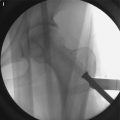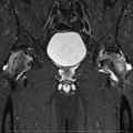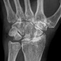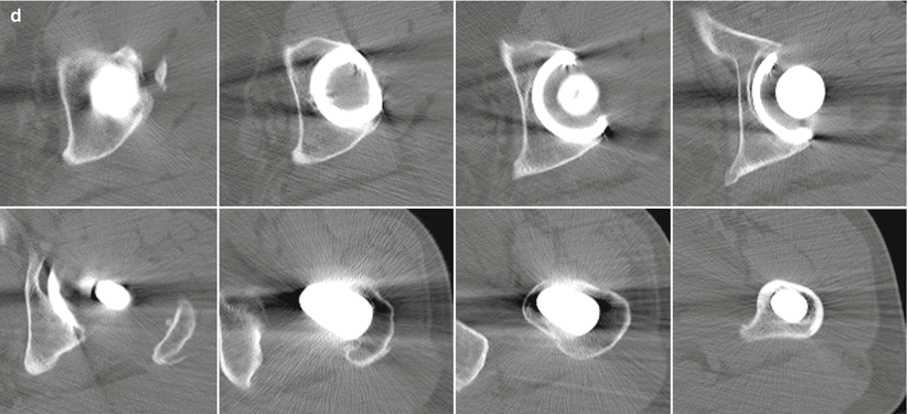
Fig. 50.1
Radiographic and computed tomographic evaluation of osteolysis of left hip of a 46-year-old man with osteonecrosis of femoral head. (a) An anteroposterior view of left hip before the operation shows sclerotic lesion in the femoral head. (b) An anteroposterior view of the left hip made 7 days after surgery reveals that Duraloc 100 series cementless acetabular component and IPS femoral component are fixed in a satisfactory position. (c) An anteroposterior view of the left hip made 12 years after surgery shows that acetabular and femoral component are fixed in a satisfactory position. Calcar round off is observed. There is no evidence of radiolucent line, polyethylene wear, or osteolysis around the acetabular or femoral component. (d) Computed tomographic scanning of left hip taken 12 years after the surgery reveals no evidence of osteolysis around the acetabular or femoral components
Two hips (1.5 %) were dislocated 3 and 5 days, respectively, after the operation and were treated successfully with a closed reduction and an abduction brace for 3 months. No further dislocation was observed in these hips until 11.6-year follow-up. No hip had a revision or aseptic loosening of acetabular and/or femoral component. Kaplan-Meier survival analysis, with revision as the end point for failure, showed that the rates of survival of both acetabular and femoral components at 11.6 years were 100 % (95 % confidence interval, 98–100).
50.4 Discussion
Survivorship of total hip arthroplasty in young patients is poorer than in older patients. Young patients may have acquired hip disease from a multitude of different causes and have different activity levels. The majority of patients in our series continue to participate in high-demand activities, including moderate to heavy manual labor. These disease entities (osteonecrosis) and the level of activity did not seem to affect the longevity of fixation of acetabular and femoral components in these young cohorts. We believe that several factors were responsible for our good results: improved design (the proximal canal-fitting design of the femoral stem including pronounced lateral flare, anteroposterior buildup, and short and narrow polished distal stem) and surgical technique for implantation of the cementless stem, the strong trabecular bone in young patients, utilization of an alumina ceramic femoral head with HXLPE liner, and small and light patients.
Our results are consistent with other studies [20, 25, 28–36]. In one study with a minimum 5-year follow-up, the wear rate of the HXLPE liner was 0.029 mm per year and 0.065 mm per year for conventional polyethylene [29]. In another study with a minimum 7-year follow-up, the mean steady-state wear rate of highly cross-lined polyethylene was 0.005 mm per year (95 % confidence interval, ±0.15 mm per year) [35]. McCalden et al. [36] reported that the mean femoral head penetration rate in the first through fifth years in patients treated with the HXLPE was 0.003 mm per year (95 % confidence interval, ±0.027 mm per year). Using edge-detection techniques at 4 years, the rate for HXLPE wear was 0.007 mm per year and 0.174 mm per year for conventional polyethylene [31, 32].
Bitsch et al. [32] reported, after a mean follow-up of 5.8 years (range, 5.0–7.7 years), that the mean femoral head penetration was 0.031 mm per year (range, 0.04–0.196 mm per year) in hips with a Marathon HXLPE (DePuy, Warsaw, Indiana) and 0.104 mm per year (range 0.04–0.196 mm per year) in hips with an Enduron polyethylene liner (DePuy). Osteolysis was not observed in any of hips with a Marathon HXLPE. Engh et al. [33] reported a reduction in the mean wear rate for Marathon HXLPE of 95 % (0.01 ± 0.12 mm per year) compared with Enduron liners (0.19 ± 0.12 mm per year). Kim et al. [35] observed that the mean Marathon HXLPE penetration rate was 0.05 ± 0.02 mm per year and no hip had aseptic loosening or osteolysis in young patients with femoral head osteonecrosis. In all but four patients in our study, the penetration rate of Marathon HXLPE was below the osteolysis threshold (0.1 mm per year). No detectable osteolysis using plain x-ray and computed tomography was observed in any hip in our study, which is consistent with other studies [29, 32, 33].
The first generation of HXLPE had documented reductions in fatigue, tensile, and toughness properties [30]. In the current series, no hip had polyethylene liner fracture. We believe that the use of adequate thickness of acetabular polyethylene liner (thicker than 6 mm) and satisfactory position of acetabular component led to absence of polyethylene liner fracture.
There has been some concern that smaller wear particles are produced with HXLPE than with conventional polyethylene, leading to a higher functional biologic activity [37, 38]. However, in our 11.6-year follow-up data, no evidence of acetabular or femoral osteolysis was observed. A longer follow-up is necessary to make conclusions about the biologic activity of HXLPE.
While there is a risk of ceramic femoral head fracture, the risk was low enough to go undetected in our small series and was outweighed by the excellent 11.6-year outcomes in a highly active, young group of patients.
There are some limitations of this study. First, the hip scoring system and the measurement of polyethylene wear are prone to interobserver variability. However, the chance-corrected kappa coefficient that was calculated to determine interobserver agreement of hip scoring and wear measurement was between 0.78 and 0.85. Second, there is some potential for bias as this is one technique and does not have a control or comparison group. Finally, the follow-up was not long enough to make conclusions regarding the theoretical advantage of alumina ceramic-on-HXLPE bearing. Strengths of this study are completeness and length of follow-up as well as consistency of clinical and radiographic examinations.
Our data suggest that the current generation of cementless acetabular and femoral components with alumina ceramic-on-HXLPE bearing has been functioning well, with no osteolysis at a 10.5-year minimum and average of 11.6-year follow-up in young patients with osteonecrosis of femoral head. While the long-term survival of implants and prevalence of HXLPE wear and osteolysis remain unknown, the midterm data are promising.
References
1.
Kim Y-H, Kook H-K, Kim J-S. Total hip replacement with a cementless acetabular component and a cemented femoral component in patients younger than fifty years of age. J Bone Joint Surg Am. 2002;84(5):770–4.PubMed
2.
Kim Y-H, Oh S-H, Kim J-S, Koo K-H. Contemporary total hip arthroplasty with and without cement in patients with osteonecrosis of the femoral head. J Bone Joint Surg Am. 2003;85(4):675–81.PubMed
3.
Kim Y-H, Oh S-H, Kim J-S. Primary total hip arthroplasty with a second-generation cementless total hip prosthesis in patients younger than fifty years of age. J Bone Joint Surg Am. 2003;85(1):109–14.PubMed
