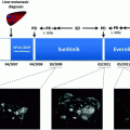
Fig. 9.1
PNET angiogenesis. Angiogenesis in PNET is primarily made from endothelial cells covered by pericytes. Endothelial cells are dependent on VEGF and VEGFR for maintenance and survival. Pericytes are dependent on PDGF and PDGFR. Sunitinib is a tyrosine kinase inhibitor that inhibits VEGFR and PDGFR, inhibiting endothelial cells and pericytes survival
Laboratory results and clinical data from phase I/II clinical trials showed evidence, suggesting that antiangiogenic agents may have potent activity in patients with advanced PNETs. In this chapter, we will review the biology of PNETs along with mechanisms yielding to the development of tumor angiogenesis. We will focus on the results obtained with sunitinib in patients with well-differentiated PNET.
Characteristics of Angiogenesis in PNETs
Well-differentiated PNETs are often characterized by the abundance of tumor vessels. Contrasting with angiogenesis in other tumor types such as glioblastomas and colorectal and breast carcinomas, in which angiogenesis is associated with a poor prognosis, angiogenesis in well-differentiated PNETs is eventually associated with a good prognosis [17]. Tumor differentiation and angiogenesis (as measured by CD31 staining) in pathological specimens are often correlated with radiological features using CT scan. Paradoxically, well-differentiated tumors are far more angiogenic than poorly differentiated carcinomas. Consistently, vascular density is correlated with tumor enhancement at the pancreatic phase of the CT scan. In univariate analysis, high microvessel density (immunostaining) and vascular enhancement in the tumors in CT scan have been associated with a favorable survival [18]. In addition, Couvelard et al. [19] have reported that high microvascular density was prevalent in well-differentiated PNETs compared to poorly differentiated carcinomas that displayed fewer vessels. Moreover, well-differentiated tumors usually have high cytoplasmic expression of VEGF and HIF-1α. In contrast, poorly differentiated carcinomas are associated with nuclear HIF-1α and high membrane expression of carbonic anhydrase-9 (CA-9). Low microvascular density and high CA-9 membrane expression have been associated with a significantly poorer survival.
Data from pathological series have elucidated biological features characterizing subgroups of sporadic PNETs. In the literature, somatic mutations of VHL were considered to represent a rare event in sporadic PNETs [20, 21] as compared to hereditary form of PNET. However, Schmitt et al. [22] recently reported that up to 25 % of sporadic PNETs display alterations in the VHL expression. Importantly, in this study performed mainly in well-differentiated PNETs, 14/78 cases (18 %) had deletion of the VHL gene (FISH), while 2/35 (6 %) cases showed methylation of the VHL promoter region. Interestingly, these genomic abnormalities were associated with low expression of VHL RNA in 25 % (8/32 cases). Consistently, approximately one-third of tumor samples showed positive staining of hypoxia target proteins including HIF-1α, CA-9, and GLUT-1 in 29, 44, and 34 % of PNET, respectively. Correlating VHL alterations and hypoxia with survival parameters, the authors observed that both VHL mutation and positive CA-9 expression were associated with poor outcome [22]. Furthermore, evidence has suggested that the expression of other proteins acting as oxygen sensors in cells such as prolyl hydroxylase domain proteins (PHD)-1, PHD-2, and PHD-3 was also correlated with a poor outcome such as tumor metastases, tumor recurrence, and lower survival [23]. Changes in metabolism and chromatin remodeling were recently observed in genetic investigations, identifying that DAXX/ATRX and MEN1, along with genetic disorders in the mTOR pathway, are frequently observed in PNETs [24]. Sporadic PNETs with adverse outcome remain good clinical models for therapeutic approaches targeting hypoxia-associated angiogenesis (HIF-1α-dependent VEGFR activation) and/or mTOR signaling pathway.
Studies of Angiogenesis in RIP-TAG Transgenic Mice
For three decades, Hanahan et al. have contributed to the field of endocrine malignant tumors by developing in 1985 a mouse model named RIP1-Tag2 (RT2). In this model, the insulin promoter was coupled with the large-T antigen of SV40, yielding transgenic mice that develop islet cell dysplasia, in situ carcinoma that could subsequently evolve according to a comprehensive multistage carcinogenesis mimicking human PNETs [25]. Unlike xenograft models transplanted in immune-deficient mice in which the murine stroma participate in tumor angiogenesis, the RT2 transgenic model offers the opportunity to explore tumor cells within their own physiological environment and angiogenesis. The RT2 model appears particularly important to explore interactions between cancer cells and “non-tumor” stromal cells, such as endothelial cells and pericytes that are responsible for the development of tumor angiogenesis. This model is also recognized as a prototype model for stepwise processes of tumor development and progression via multiple stages. For instance, Langerhans islet cells display stages of multifocal carcinogenesis such as in situ carcinoma, eventually undergoing the angiogenic switch that results in invasive, bulky, and potentially lethal endocrine carcinoma.
Multiple signaling pathways related to IGF/IGF-1R, MMP-9 and MMP-2, VEGF-A/VEGFR2, mTOR, EGFR, and PDGF-B/PDGFRβ [12, 26–28] are frequently activated in several cell types such as endothelial and tumor cells, pericytes, and stromal cells in PNETs [27–29]. The RT2 model appears highly reproducible and has been frequently used as a tool to explore pharmacological strategies targeting different stages of PNET carcinogenesis [29–31].
However, the RT2 model also suffers limitations. For instance, the incidence of tumor necrosis and hypoxia-related signaling activation remains limited in RT2 as compared to human PNETs. Another limiting factor is the low incidence of metastases occurring at late stages of RT2, as most of the mice may die at advanced stages from tumor-induced hypoglycemia before developing distant metastases. This limitation was addressed by developing a variant model of RT2 that consisted of a double transgenic RIP1-Tag2 and RIP7-Igf-1R mouse model, overexpressing the type I insulin-like growth factor receptor (IGF-1R) in pancreatic islets [26]. This model yields to a more invasive and metastatic phenotype. Although the life span of mice in this later model is reduced compared to RT2 counterparts, this model has been considered to be more relevant to study PNET metastases as it reproduces features closer to advanced human PNETs.
Evaluation of Antiangiogenic Agents in the RT2 Model
Targeted agents including antiangiogenic agents have been explored using the RT2 model (Table 9.1). Initially, preclinical evaluations have focused on matrix metalloproteinase (MMP) inhibitors, angiostatin, and endostatin. Preclinical evaluations using [29, 30] these compounds failed to translate into clinical benefits due to poor pharmacokinetic parameters (angiostatin, endostatin) or unexpected toxicities (MMP inhibitors). In recent years, new compounds, including tyrosine kinase inhibitors against VEGFR and PDGFR and mTOR inhibitors, have been extensively investigated. VEGFR2 inhibitors and mTOR inhibitors were able to interfere efficiently with angiogenesis and tumor development in RT2 models [31]. VEGFR2 inhibition almost completely prevented dysplastic lesions to undergo the angiogenic switch in prevention experiments and also significantly reduced the size of tumors in intervention and regression experiments. The antitumor effects of antiangiogenic agents and mTOR inhibitors were associated with a significant reduction in vessel density and permeability. Immunohistochemistry studies revealed that rapamycin also increased significantly the frequency of apoptotic cancer cells [31]. The promising results reported in these experiments are consistent with the recent clinical trials using sunitinib and everolimus that were recently conducted in patients with PNET [32, 33].
Table 9.1
All-grade emergent adverse events (%) occurring in ≥20 % of patients in either sunitinib or placebo arm in the sunitinib phase III trial [32]
Sunitinib (n = 83) | Placebo (n = 82) | |
|---|---|---|
Diarrhea | 49 (59) | 32 (39) |
Nausea | 37 (45) | 24 (29) |
Asthenia | 28 (34) | 22 (27) |
Vomiting | 28 (34) | 25 (30) |
Fatigue | 27 (32) | 22 (27) |
Hair color changes | 24 (29) | 1 (1) |
Neutropenia | 24 (29) | 3 (4) |
Abdominal pain | 23 (28) | 26 (32) |
Hypertension | 22 (26) | 4 (5) |
Hand-foot syndrome | 19 (23) | 2 (2) |
Anorexia | 18 (22) | 17 (21) |
Stomatitis | 18 (22) | 2 (2) |
Taste disturbances | 17 (20) | 4 (5) |
Epistaxis | 17 (20) | 4 (5) |
Understanding Resistance to VEGFR Inhibitors in RT2 Mice
Emerging resistance to VEGFR inhibitors appears as an important issue in clinical trials that remains poorly understood. Several studies using the RT2 model have noticed that continuous exposure to antiangiogenic agents such as VEGFR inhibitors may eventually be associated with the emergence of acquired resistance. Casanovas et al. [34] have reported that short-term treatment with VEGFR2-blocking antibodies resulted in smaller tumors. However, more prolonged exposures that were associated with initial tumor shrinkage were subsequently followed by a tumor regrowth mimicking tumor progression occurring in patients with acquired resistance to antiangiogenic agents. When regrowing, tumors were shown harboring an invasive phenotype. Islet tumor cells that developed under VEGFR2 therapies were more prompt to invade surrounding exocrine pancreatic tissues and induce capsule breakage. Despite a sustained inhibition of VEGFR2, most tumors analyzed at the time of progression still displayed a high microvascular density with leaky vessels and microvascular hemorrhages comparable to untreated tumors. This suggested that tumor angiogenesis might have developed using VEGFR2-independent mechanisms at the time of tumor resistance. In this paper, the authors pointed out the important role of hypoxia and HIF-1α in resistant tumors. Further analysis revealed that several proangiogenic factors, including fibroblast growth factors (FGFs), ephrins, and angiopoietins, were upregulated in resistant tumors. Interestingly, these proangiogenic factors were mainly upregulated in tumor cells compared to endothelial cells. These data suggest that hypoxia triggered by VEGFR2 inhibitors may induce multiple alternative factors of angiogenesis and facilitate a transition toward a more invasive phenotype. Consistently, our team also reported such an induced epithelial-to-mesenchymal transition in patients receiving long duration of treatment with sunitinib in patients with hepatocellular carcinomas [35]. More recently, data have also suggested that resistance to VEGFR inhibitors may be associated with c-MET activation in part explaining the reasons why tumor progression in PNETs may be associated with the development of an invasive and metastatic phenotype.
Sunitinib as a Prototype Drug-Inhibiting Angiogenesis in PNET
Sunitinib malate (SUTENT®; Pfizer Inc, NY, USA) is an antiangiogenic agent initially approved for the treatment for advanced renal cell carcinoma and imatinib-resistant/imatinib-intolerant gastrointestinal stromal tumors (GIST). Sunitinib inhibits VEGFR-1, VEGFR-2, and VEGFR-3, PDGFR-α and PDGFR-β, and c-KIT, in addition to FMS-like tyrosine kinase 3 (FLT3), colony-stimulating factor-1 receptor (CSF-1R), and glial cell line-derived neurotrophic factor receptor (rearranged during transfection (RET)) [36]. The above-mentioned kinase receptors have been described in NETs including PNETs, providing a rationale for the clinical evaluation of sunitinib in PNET [12, 28]. Furthermore, as a result of inhibition of VEGFR and PDGFR, data on sunitinib in the RT2 mouse model demonstrated a significant reduction in endothelial cells and pericyte coverage of tumor vessels [37]. Experiments performed in this model showed that the inhibition of either PDGFR or VEGFR might inhibit the growth of tumors by preventing the malignant transformation and by acting on established tumors. The magnitude of the effects was greater when both receptors were inhibited together than either VEGFR or PDGFR alone, suggesting a potential for greater therapeutic benefit when both of the receptor families were inhibited concurrently. As a result, therapeutic experiments using sunitinib in mice with established RT2 tumors demonstrated significant reduction in tumor size and extended survival in mice treated with sunitinib as compared to a saline placebo, establishing the relevance of this model for clinical applications [38].
Activity of Sunitinib in Phase I–II Clinical Trials
In the first-in-man phase I trial with sunitinib, potent antitumor activity (unusually high number of objective radiological responses) was observed in several tumor types [39]. Patients who were enrolled in the phase I trial presented tumors that expressed VEGFR, PDGFR, and c-KIT, that were highly angiogenic, and that were resistant to cytotoxic agents. Objective responses were observed in renal cell carcinoma and imatinib-resistant GIST, leading to phase II/III trials that subsequently demonstrated the efficacy of sunitinib in those two indications. Three patients with NETs entered in the phase I trial. These patients were primarily referred for tumor progression after several lines of chemotherapy. One patient experienced a prolonged partial response, and two patients achieved sustained minor responses [39]. Subsequently, a multicentre phase II trial was launched with sunitinib (50 mg/day 4 weeks on/2 weeks off) in patients with carcinoids and PNETs [40]. Among 66 patients with advanced PNETs (28.8 % of patients with functioning tumors), the objective response rate was 16.7 % with 56.1 % of patients experiencing tumor stabilization for more than 6 months, leading to a median time-to-tumor progression of 7.7 months. Patient-reported outcome data in this phase II trial showed no detrimental effects of sunitinib on quality of life across repeated cycles.
Dataset to Evaluate the Efficacy of Sunitinib in PNET
The efficacy of sunitinib in PNET was demonstrated in a large placebo-controlled multicenter phase III trial. In this study [32], sunitinib was given continuously at the daily dose of 37.5 mg (with no dose interruption). The continuous daily dosing yields same dose intensity than the 50 mg/day dosing given for four weeks every six weeks. This dosing was selected to avoid dose interruption, potential flair-up in tumor angiogenesis following treatment discontinuation, and was though to induce less acute, i.e., more manageable side effects in a patient population expected to receive sunitinib over a very long period of time. The study was designed to detect a 50 % improvement in the progression-free survival (PFS) in patients receiving sunitinib compared to placebo, expecting that placebo-treated patients would achieve a median PFS of less than 6 months. Based on theses assumptions, 340 patients would have been required to complete this trial. Since PFS was the primary end point in this trial, patients were allowed to crossover to sunitinib at the time of tumor progression in open-label sunitinib continuation studies. This study was terminated earlier than anticipated based on advice provided by an independent data monitoring committee who accessed unblinded data. This committee detected a higher occurrence of deaths and serious adverse events in patients receiving placebo. The committee also identify that at the time of their evaluation, the PFS in patients receiving sunitinib was double of that in the placebo group. Despite a relatively low number of events, this observation of 100 % improvement in PFS made unlikely that the trial would have been missing its primary end point of 50 % improvement in PFS at the time of the formal primary analysis. Furthermore, it was considered unethical to continue randomizing patients in the placebo group, which was resulting in a greater risk of early death and adverse events. Therefore, the recommendation was made to discontinue randomizing patients and give a chance to patients previously randomized to placebo to access open-label sunitinib. This recommendation was subject to criticism. It was primarily argued that this early termination, occurring before the expected number of events, might have reduced the power of the study and overestimated the magnitude of PFS benefit of sunitinib in this patient population. However, the PFS benefit (as reflected by the hazard ratio) in this study was very high and has been correlating with a high tumor control rate and an improved overall survival. Those benefits observed on secondary end points provided additional supportive evidence of the drug efficacy and easily set aside most of reservations based predominantly on pure statistical considerations.
Related posts:
 New Anticancer Agents in Neuroendocrine Tumors
Inhibition of mTOR in Neuroendocrine Neoplasms of the Digestive Tract
Clinical Management of Targeted Therapies in Neuroendocrine Tumours
New Anticancer Agents in Neuroendocrine Tumors
Inhibition of mTOR in Neuroendocrine Neoplasms of the Digestive Tract
Clinical Management of Targeted Therapies in Neuroendocrine Tumours
 Overcoming Resistance to Targeted Therapies: The Next Challenge in Pancreatic Neuroendocrine Tumors (PNETs) Treatment
 Measuring the Relationship of Quality of Life and Health Status: Including Tumor Burden, Symptoms, and Biochemical Measures in Patients with Neuroendocrine Tumors
 Imaging of Neuroendocrine Tumors and Challenges in Response Evaluation for Targeted Therapies
Stay updated, free articles. Join our Telegram channel

Full access? Get Clinical Tree


