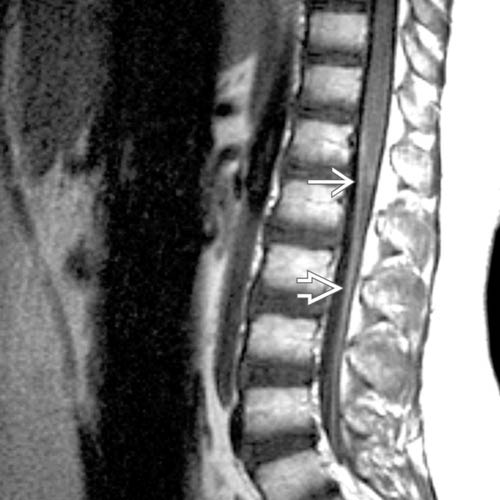
(Left) Sagittal T1WI MR in a leukemia patient with leg weakness following intrathecal chemotherapy shows normal appearance of the conus and cauda equina.
(Right) Sagittal T1WI C+ MR in a leukemia patient with leg weakness following intrathecal chemotherapy reveals abnormal smooth, thin, linear enhancement of the ventral conus pia and cauda equina .
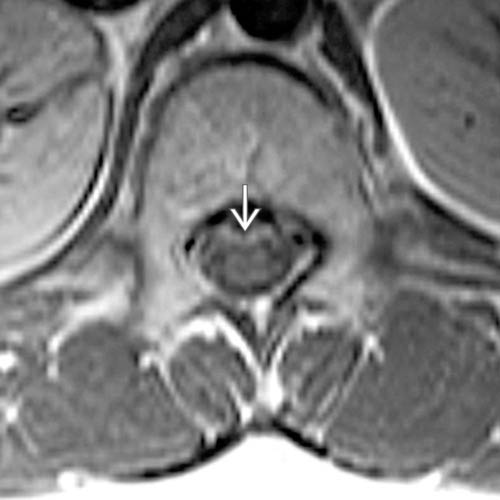
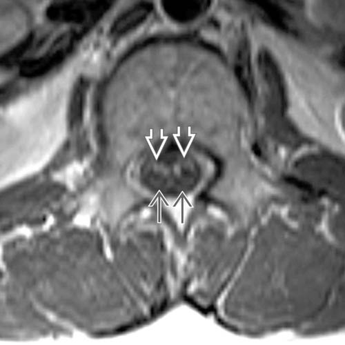
(Left) Axial T1WI C+ MR through the conus medullaris confirms smooth, thin, linear enhancement of the ventral conus pia and cauda equina .
(Right) At the level of the cauda equina, axial T1WI C+ MR demonstrates characteristic smooth thin linear enhancement of the ventral cauda equina nerve roots but not the dorsal cauda equina nerve roots .
 and cauda equina
  .
.
 .
.
 but not the dorsal cauda equina nerve roots
  .
.