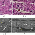
Fig. 31.1
Applications of ICG fluorescence imaging during lung metastasectomy. Metastatic lesions with diameters of 5 mm (a), 1 mm (b), and 0.07 mm (c) were detected by ICG fluorescence imaging. Initial photos (a1, b1, c1) and following photos (a2, b2, c2) indicate the same view of each lesion under room light and ICG fluorescence imaging, respectively. Histopathological finding of b1 lesion appears in b3
31.2.2.2 Lymph Node Metastases
Although infrequent, the ICG method is effective in treating lymph node metastases. Metastatic lymph nodes of the hepatic portal region and mediastinum can be confirmed with satisfactory contrast. This method is particularly effective in cases where the involved lymph node is situated among several other lymph nodes, as the metastatic lymph nodes can be singled out and resected (Fig. 31.2).


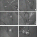
Fig. 31.2
Intraoperative view of a metastatic mediastinal lymph node. (a) ICG fluorescence imaging indicates the node. (b) Room light view of the lesion. (c) After an incision of the mediastinal pleura
31.2.2.3 Peritoneal Metastases
In cases where metastases are suspected in the peritoneum, a laparotomy is performed, and the ICG method is used (Fig. 31.3). In such cases, the timing of ICG administration is approximately 72 h before surgery. The reason for this is that the florescence of the digestive tract is too strong due to excreted bile.
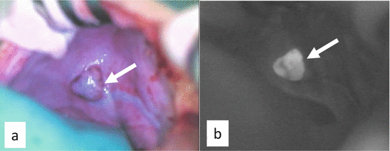

Fig. 31.3
Intraoperative view of a metastatic peritoneal lesion of the right diaphragm. (a) Room light view of the lesion. (b) ICG fluorescence imaging
31.3 Problems
31.3.1 False-Positive
Of the 250 masses that were identified as positive and resected, tumors could not be histopathologically detected in 29 masses. Interestingly, these false-positives were concentrated in two patients and were not recognized in the remaining eight. The following two reasons may be possible explanations for these false-positives:
1.
Fluorescence of tissue other than the tumor. According to observations through a fluorescence microscope within the fluorescence wavelength of ICG, we were able to confirm that thrombi and granuloma were the cause of this non-tumorous fluorescence. While it is unclear why ICG detected these “masses,” they consisted of tissue not observed in healthy lungs and may be an alteration after chemotherapy.
2.
The possibility that the tumor is too small to be detected. The smallest tumor histopathologically confirmed with the ICG method was 62 μm. On the other hand, paraffin-embedded tissue sections are usually cut out at approximately 20 μm. Therefore, if there are tumors smaller than 20 μm, it is possible that these would not be included in the slice.
Stay updated, free articles. Join our Telegram channel

Full access? Get Clinical Tree


