We discuss the arterial supply of the cranial nerves from their exit out of the brain stem to their exit from the skull base. Four distinct groups can be differentiated from an embryologic and phylogenetic standpoint. Understanding the arterial supply to the cranial nerves and the potential anastomoses is paramount in the endovascular treatment of dural AV shunts and highly vascularized tumors of the skull base to avoid neurologic deficits.
Most cranial nerves are peripheral nerves. They originate from one or multiple brain stem nuclei and course through the skull base to innervate the orbit and derivatives from the six branchial arches mostly within the head and neck region. Understanding of the arterial vascularization of the cranial nerves is derived from anatomic, microsurgical, and angiographical studies. Under normal conditions, the caliber of these arteries, is small, ie, between 100 and 300 microns. Thus they can be visualized during digital subtraction angiography (DSA) only with superselective injections. Although noninvasive imaging methods have improved considerably and are able to demonstrate the course of the cranial nerves, they remain insufficient to detect their arterial vascularization.
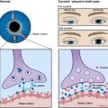
Few pathologies may lead to specific involvement of the vascularization of the cranial nerves. Diabetes or viral infections may be the cause of vasculitis of these small vessels and subsequent nerve ischemia and deficit. This is described for example in Bell’s palsy where the viral involvement is responsible for a breakdown of the blood-nerve barrier resulting in gadolinium enhancement on MRI.
Some pathologic processes induce enlargement of dural arteries and subsequently of arteries to the cranial nerves without compromising their supply: these encompass highly vascularized and/or dural tumors (paragangliomas, juvenile angiofibromas, meningiomas) or dural arteriovenous shunts. The endovascular treatment of these pathologies requires a careful understanding of the regional anatomy and their vascular source as well as possible dangerous anastomoses between the cranial nerve supply and the central nervous system to avoid complications.
The phylogeny and neuroembryology remain essential to understand anatomic variations that may be encountered. At the spinal level every metameric artery is supplying its corresponding nerve root, from its origin at the spinal cord to the foramen. The metameric distribution, although less obvious, will help to understand the vascularization of the cranial nerves at the cervicocephalic level.
The aim of this chapter is to discuss the arterial supply of the cranial nerves from their exit out of the brain stem to their exit from the cranial cavity. The dural vascular network and its anastomoses between external carotid, internal carotid, and vertebral artery through the respective foramina of the skull base are also considered. Outside the cranial cavity, the arterial nerve root supply is derived from other branches of the external carotid artery and will not be further detailed. Being outpouchings of the brain rather than true cranial nerves, the first and second nerves are not considered as peripheral nerves and for didactic and practical reasons are not considered in this text. Briefly, the first cranial nerve is supplied by the olfactory artery and ethmoidal branches of the ophthalmic artery. The optic nerve is supplied by the proximal ophthalmic artery as a remnant of the ventral and dorsal ophthalmic arteries.
For better comprehension of embryology and anatomic correlations, in the following description, the cranial nerves were grouped as belonging to different embryonic systems. For each group, the embryologic and phylogenic part is described after brief overviews of the cranial nerve anatomy and their respective arterial descriptive anatomies are given.
Group of extraocular nerves and ophthalmic root of the trigeminal nerve: III, IV, VI, and V1
Cranial Nerves: Descriptive Anatomy
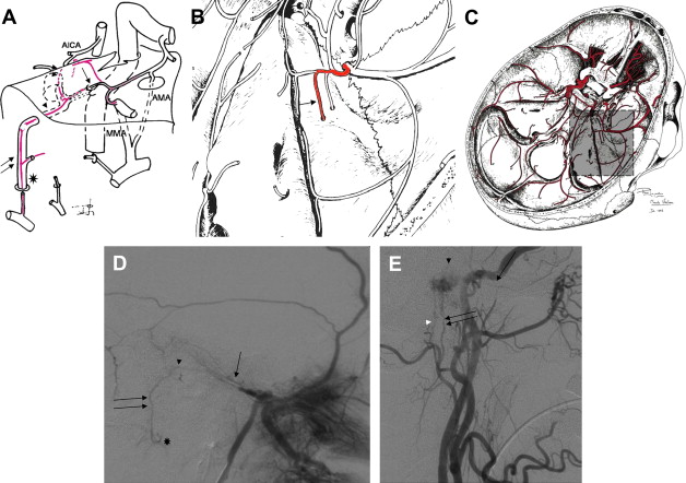
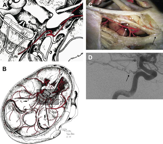
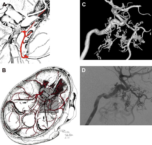
The cranial nerves of this group emerge from the brain stem at different points, course into the subarachnoid cisterns of the skull base and posterior fossa, to enter subsequently into the cavernous sinus ( Figs. 1 and 2 ). The third nerve is exiting the brain from the medial sulcus at the medial surface of the cerebral peduncle and courses through the interpeduncular fossa between the anterosuperior cerebellar artery and the posterior cerebral artery. It passes over the basal attachment of the tentorium to the posterior part of the roof of the cavernous sinus (oculomotor triangle). After penetrating the roof of the sinus, the oculomotor nerve courses inside its lateral wall passing forward through the superior orbital fissure to innervate intraorbital structures.
The fourth nerve originates from a controlateral nuclei and is the only one to emerge from the dorsal surface of the brain stem. It turns around the cerebral peduncle and courses anteriorly, within the cerebellomesencephalic, quadrigeminal, and ambient cisterns, parallel to the anterosuperior cerebellar and posterior cerebral artery and basal vein until it enters the cavernous sinus, just lateral to the oculomotor nerve. The trochlear nerve runs inside the inner layer of the lateral wall of the cavernous sinus, below the third nerve to leave this region via the superior orbital fissure.
The sixth nerve arises from the lower portion of the pons and courses along the cerebellopontine angle’s cistern to reach Dorello’s canal or petroclival venous confluence at the apex of the petrous bone. Inside the cavernous sinus, the nerve runs in its lower part, on the lateral surface of the internal carotid artery (ICA) and below the inferolateral trunk until it enters the superior orbital fissure in its medial aspect.
Arterial Descriptive Anatomy
Contribution from the vertebrobasilar system to the proximal cisternal course of the cranial nerves
In their cisternal course, after they emerge from the brain stem, nerves III, IV, and VI receive arterial supply from branches of the vertebrobasilar system. The third nerve may receive arterial supply in the vicinity of the posterior perforating substance from the basilar artery or from the posterior cerebral artery; subsequently this arterial supply may also concern parts of the thalamus. There are either two lateral arteries or one common trunk. The fourth nerve is supplied by branches from the anterosuperior cerebellar artery or from circumferential arteries of the P1 segment of the posterior cerebral artery.
Contribution from dural and transosseous arteries
The dural arteries contribute to the supply of the cranial nerves in their dural and transosseous course. Nerves III and IV nerves are supplied by the marginal artery of the tentorium cerebelli on the roof of the cavernous sinus. The marginal artery of the tentorium is an arcade between orbital vessels and branches of the carotid siphon. Many anatomic variations may be observed in this region. The artery may arise from the middle meningeal artery, the ophthalmic or lacrimal artery, the inferolateral trunk, or the posterior group of collaterals of the internal carotid artery.
The sixth nerve is supplied by a clival network along its course anterior to the pons. Medial and lateral clival arteries are caudally fed by the hypoglossal and jugular branch of the neuromeningeal trunk of the ascending pharyngeal artery. Cranially, the medial and lateral clival arteries arising from branches of the ascending segment of the carotid siphon supply this nerve.
The anteromedial branch of the inferolateral trunk is supplying nerves III, IV, VI, and V1 in the cavernous sinus and the superior orbital fissure.
Arterial Embryology and Phylogeny
This group of cranial nerves (III, IV, VI, and V1) is under the control of the dorsal ophthalmic artery, whose vestige is the inferolateral trunk of the carotid siphon. The metameric structures they are linked to are the prebranchial somites. The medial clival artery and the posteroinferior hypophyseal trunk belong to the remnant of the primitive maxillary artery. The lateral clival artery is the remnant of the trigeminal artery.
Group of trigeminal ganglion and trigeminal nerves V2, V3, and Vm, and facial nerve VII and vestibulocochlear nerve VIII
Trigeminal Ganglion and Trigeminal Nerves V2, V3, and Vm
Cranial nerve descriptive anatomy
The fifth nerve emerges from the lateral surface of the pons (see Fig. 2 ; Fig. 3 ). Both the sensory roots and the motor portion cross the subarachnoid space below the superior petrosal sinus to reach the upper edge of the petrous bone at the level of Meckel’s cave and the trigeminal ganglion (gasserian ganglion). The motor and sensory roots are regrouped and diverged into the three major roots, ophthalmic (V1), maxillary (V2) and mandibular nerves (V3), to reach their foramina as indicated in ( Table 1 ).
| Intracranial Feeders | ||||
|---|---|---|---|---|
| Cranial Nerves | Intracisternal Vertebrobasilar Origin | Epidural Internal Carotid Artery (ICA) Origin | Skull Base Foramen | External Carotid Arteries Feeders |
| III | PCA |
| Superior orbital fissure | Ophthalmic, or lacrimal, or MMA tentorial branches |
| IV | ASCA |
| Superior orbital fissure | Ophthalmic, or lacrimal, or MMA tentorial branches |
| V 1 | Remnant of the trigeminal artery |
| Superior orbital fissure | Ophthalmic, or lacrimal, or MMA tentorial branches |
| V 2 | Remnant of the trigeminal artery | Medial branch of the ILT to the artery of the foramen rotundum | Foramen rotundum | Artery of the foramen rotundum (distal maxillar artery) |
| V 3 and V m | Remnant of the trigeminal artery | Posterior branch of the ILT, to the accessory meningeal artery | Foramen ovale | Accessory meningeal artery (proximal maxillar artery) |
| VI |
| Superior orbital fissure | Clival arteries from neuromeningeal trunk of APA | |
| VII | Auditory artery, branch of the AICA |
|
| |
| VIII | Auditory artery, branch of the AICA | |||
| IX | Vertebral artery |
| Neuromeningeal trunk of APA (jugular branch) | |
| X | Vertebral artery |
| Neuromeningeal trunk of APA (jugular branch) | |
| XI | Vertebral artery |
| Neuromeningeal trunk of APA (jugular branch) | |
| XII | Vertebral artery | Hypoglossal canal | Neuromeningeal trunk of APA (hypoglossal branch) | |
Arterial descriptive anatomy
Contribution from the vertebrobasilar system to the proximal cisternal course of the cranial nerves
The trigeminal nerve in its proximal cisternal course, after emerging from the pons, is supplied by the vestigial artery of the trigeminal artery that arises from the basilar artery.
Contribution from dural and transosseous arteries
The trigeminal ganglion in Meckel’s cave is supplied by the lateral artery of the trigeminal ganglion that arises from the vertical portion of the intracavernous ICA, and by cavernous branches from the middle meningeal artery. V2 is supplied by the artery of the foramen rotundum, which is fed by the inferolateral trunk that arises from the cavernous horizontal segment of the internal carotid artery. The artery of the foramen rotundum courses through the foramen rotundum and anastomoses with the distal maxillary artery. V3 and Vm have a common course through the oval foramen: they are supplied by the accessory meningeal artery, which represents an anastomosis between the posterior branch of the inferolateral trunk of the carotid siphon and the proximal maxillary artery. There is a hemodynamic balance between the accessory meningeal artery and the posterior branch of the inferolateral trunk. The variation in size of the accessory meningeal artery is significant and depends on its soft tissue supply. Depending on the depth of the course of the maxillar artery, the accessory meningeal artery may originate from a common trunk with the middle meningeal artery (in case of a superficial course of the maxillar artery) or directly from the maxillar artery (in case of a deep course). V3 and Vm are also supplied by cavernous branches from the middle meningeal artery.
Vestibulocochlear Nerve and Facial Nerve
Cranial nerve descriptive anatomy
The seventh nerve includes sensory and motor fibers and emerges from the pontomedullary junction caudal to the origin of the trigeminal nerve ( Fig. 4 ). It enters the pontocerebellar cistern and courses anteriorly above the eighth nerve before entering the internal auditory canal until it reaches the facial nerve canal and soon after the geniculate ganglion. It gives off intrapetrous branches before exiting the canal at the stylomastoid foramen.