47 Arteries of the Spinal Cord
F. Goetz, A. Giesemann
The spinal cord is supplied by three longitudinal arterial trunks—the anterior spinal artery and the two posterolateral trunks. The anterior spinal artery is formed by two roots from the vertebral arteries, and anterior radicular arteries reinforce the longitudinal channel at various levels; in the cervical part the anterior radicular arteries arise from the vertebral artery, thyrocervical trunk, and costocervical trunk. Branches from intercostal and lumbar arteries join the longitudinal system at the thoracic and lumbar segments. At the conus terminalis, a periconal anastomotic circle is present connecting anterior spinal with posterior spinal arteries. Usually one artery is larger than the other, that is, the great anterior medullary artery (artery of Adamkiewicz radicularis magna), which is more often found on the left than on the right side and usually between the levels T9 and L2. Variations of the posterior radicular arteries are not shown in Fig. 47.1. The great variability in the number of radicular arteries, the prevalence of the left side in the thoracic and lumbar parts and the level of the radicular arteries has been explained by the embryological segmental blood supply and the preferential left side, by the more direct blood flow from the aorta to the left radicular arteries.1 Paraplegia following orthopaedic surgery of the spine or operation of the aorta stresses the clinical relevance of the anomalies of the spinal cord blood supply.1–16
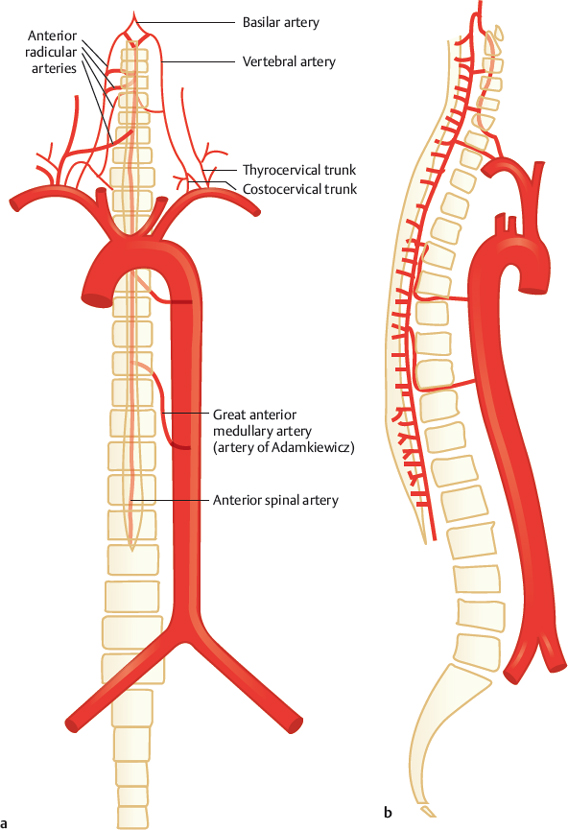
Fig. 47.1 Typical origin of the anterior radicular arteries. Schematic, anterior (a) and lateral (b) view.
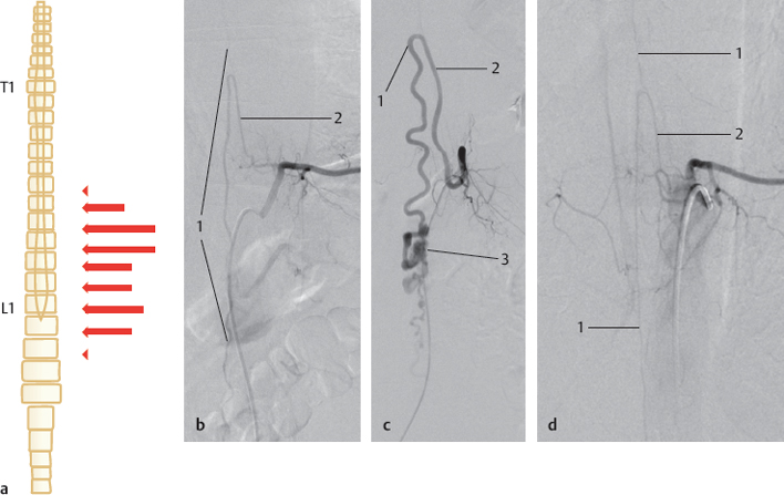
Fig. 47.2 Variable origin of the great anterior medullary artery (artery of Adamkiewicz). Schematic, anterior view (a), and X-ray angiography, anterior view, selective injection of radicular arteries (b–d). Artery of Adamkiewicz fed from T9 left (b), from T10 left in a case with a perimedullary arteriovenous malformation (c), and from L1 left (d). 1 Anterior spinal artery; 2 artery of Adamkiewicz; 3 perimedullary arteriovenous malformation. T1, first thoracic vertebra; L1, first lumbar vertebra.
Related posts:


Stay updated, free articles. Join our Telegram channel

Full access? Get Clinical Tree


