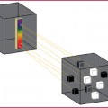
Subclavian artery (green)
Axillary artery (blue)
Brachial artery (pink)
Radial artery (yellow)
Ulnar artery (red)
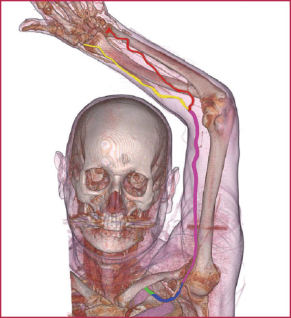
Fig. 12.1
Principal vessels of the upper extremity
Table 12.2
Collateral vessels of the subclavian artery
Vertebral artery | ||
Internal thoracic artery (Internal mammary artery) | ||
Thyrocervical trunk | Inferior thyroid artery | |
Suprascapular artery | ||
Transverse cervical artery | ||
Ascending cervical artery | ||
Superior cervical artery | ||
Costocervical trunk | Deep cervical artery | |
Superior intercostal artery | ||
Table 12.3
Collateral branches of the axillary artery
Superior thoracic artery | |
Thoraco-acromial artery | Acromial branch Clavicular branch Deltoid branch Pectoral branch |
Lateral thoracic artery | |
Subscapular artery | Thoracodorsal artery Circumflex scapular artery |
Anterior humeral circumflex artery | |
Posterior humeral circumflex artery |
Table 12.4
Collateral branches of the brachial artery
Deep artery of the arm | Medial collateral artery Radial collateral artery |
Superior ulnar collateral artery | |
Inferior ulnar collateral artery |
Table 12.5
Collateral branches of the radial artery
Radial recurrent artery | |
Palmar carpal branch | |
Superficial palmar branch | |
Dorsal carpal branch | Dorsal metacarpal arteries Dorsal digital arteries |
Princeps pollicis artery | |
Radial indicis artery | |
Deep palmar arch | Palmar metacarpal arteries Perforating branch |
Table 12.6
Collateral branches of the ulnar artery
Ulnar recurrent artery | |
Common interosseous artery | Posterior interosseous artery |
Recurrent interosseous artery | |
Anterior interosseous artery | |
Superficial palmar branch | |
Volar carpal | |
Dorsal carpal | |
Deep volar | |
Superficial volar arch | Common palmar digital arteries |
Proper palmar digital arteries |
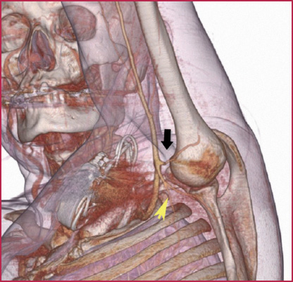
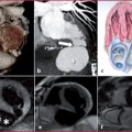
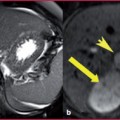
Fig. 12.2
Anterior humeral circumflex artery (arrowhead) and posterior humeral circumflex artery (arrow)
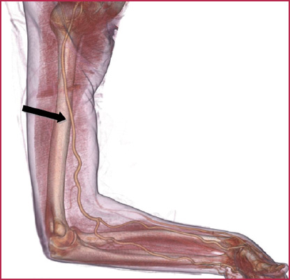
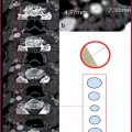
Fig. 12.3
Radial artery (arrow) and radial artery bifurcation
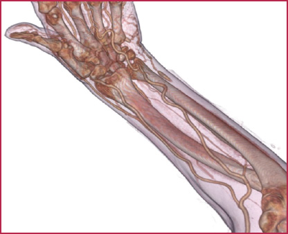
Fig. 12.4
Ulnar and radial arteries
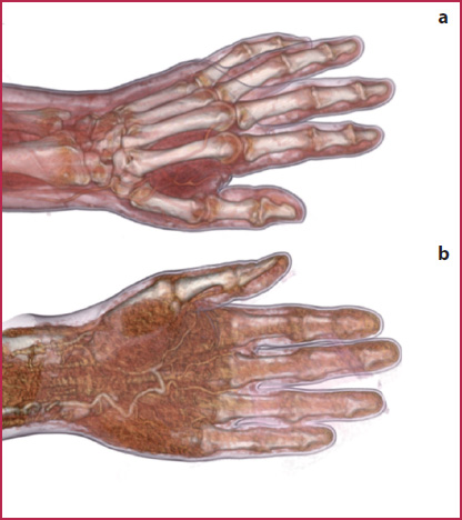
Fig. 12.5
Dorsal metacarpal arteries (a) and palmar (b)
The most frequent anatomic variants of the arteries of the upper extremity are shown in Figs. 12.6–12.8
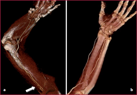
Fig. 12.6
a Early origin of the ulnar artery proximal from the proximal third of the humeral artery (arrow); b regular origin at the level of antecubital fossa
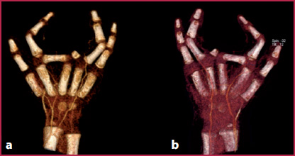
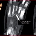
Fig. 12.7
Agenesis of the intermediate and distal phalanges of the 3rd finger. Right hand: palmar (a) and dorsal (b) surface. The ulnar and radial arteries provide branches for the 1 st finger and for the 4th and 5th fingers. The radial artery provides branches to the 3rd finger and the ulnar artery to the 1st, 2nd, 4 th and 5th fingers
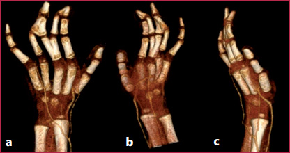
Fig. 12.8
Polydactyly with fusion of some fingers. Left hand: a dorsal surface, b palmar surface, c medial surface. The ulnar and radial arteries provide terminal branches for all the fingers, except for the 5th finger, which is vascularized by the ulnar artery
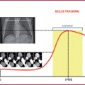
12.2 12.2 CTA Technique
The basic technical parameters and the strategies of administration of the contrast agent are reported in Tables 12.7 and 12.8. All data refer to a person with a body weight of approximately 70 kg (154 pounds). Refer to www.MDCT.net for additional information.
Patient Preparation
The patient is positioned in a prone or supine position and the head enters the scanner first with the affected extremity extended above the head, in the center of the gantry with the palm ventral and the fingers spread apart. The contralateral arm is placed at the patient’s side. Pillows or other supports are utilized as needed so that the patient can maintain this position and the extension of the upper extremity.
For the examination of the thoracic outlet syndrome (TOC), the supine patient has to be in a neutral position with the upper extremities placed along the body. The affected extremity is extended upwards with the head rotated contralaterally.
The peripheral venous access (with an 18–20 G needle) is placed in the extremity contralateral to the affected limb, in order to avoid artifacts during the passage of the contrast agent.
Remove any metallic material present in the neck, chest and upper extremities.
Table 12.7
Scan parameters
4 MDCT | 16 MDCT | 64 MDCT | 128 MDCT | Dual-energy | |
scanners | |||||
kVp | 120 | 120 | 120 | 120 | 120 |
mAs | 200-300 | 200-300 | 180-200 | 180 | 120-180 |
Collimation (mm) | 4 x 2.5 | 16 x 0.75 | 64 x 0.625 | 128 x 0.6 | 64 x 0.6 x 2 |
Slice reconstruction (mm) | 1-3 | 0.625-1 | 0.5-1 | 0.6 | 0.6 |
Slice reconstruction (mm) | 0.4 x 0.6 | 0.5-1 | 0.5 | 0.4 | 0.4 |
Table 12.8
Contrast agent injection protocol
4 MDCT | 16 MDCT | 64 MDCT |