Aorta and inferior vena cava (IVC) are frequently ipsilateral (usually right side)
• Polysplenia (PSP) syndrome: Left isomerism or bilateral left-sidedness
Usually multiple spleens, but may have single normal spleen
– Isolated reversal of splenic position (in right abdomen) common
Increased risk of complex cardiac anomalies, although less common with PSP than ASP
IVC interruption with azygos continuation very common
Bilateral bilobed lungs
Truncated/short pancreas or agenesis of dorsal pancreas
– Increased incidence of diabetes and pancreatitis
Intestinal malrotation is seen in most patients
Liver often midline with range of biliary abnormalities
Aorta usually located to left of midline
TOP DIFFERENTIAL DIAGNOSES
• Splenosis
• Accessory spleen
• Splenectomy
CLINICAL ISSUES
• ASP: Newborn or infant presentation due to cardiac disease with poor prognosis and early mortality
↑ risk of sepsis due to lack of spleen
• PSP: Infant or adult presentation with better prognosis due to lesser incidence of cardiac disease
(Left) Coronal volume-rendered CECT in a patient with polysplenia (PSP) syndrome demonstrates multiple spleens in the left upper quadrant. The multiple spleens in PSP are typically in the left abdomen, but can rarely be on the right.
(Right) Axial CECT in the same patient demonstrates a markedly dilated azygous vein .
(Left) Axial CECT in the same patient again demonstrates multiple spleens and a dilated azygous vein to the right of the aorta. Azygous continuation of the inferior vena cava (IVC) is a very common abnormality in PSP syndrome.
(Right) Axial CECT in the same patient demonstrates malrotation of the bowel, with the small bowel abnormally located in the right abdomen and the entirety of the colon in the left abdomen. Malrotation is quite common with both asplenia (ASP) and PSP syndromes.
• Complex inherited syndromes associated with absence (ASP) or multiplicity (PSP) of spleens, as well as many other anomalies
Associated Syndromes
• Heterotaxy: Abnormal embryologic placement of thoracoabdominal structures across right-left axis of body
• Situs solitus: Normal placement of thoracoabdominal organs in right-left axis
• Situs inversus: Reversal of normal positions of thoracoabdominal organs across right-left axis (mirror-image of situs solitus)
Can be subdivided into situs inversus with dextrocardia or levocardia
• Situs ambiguus (heterotaxy syndrome): Abnormal placement of thoracoabdominal structures without situs inversus
Situs ambiguus with polysplenia: Left isomerism or bilateral left-sidedness
Situs ambiguus with asplenia: Right isomerism or bilateral right-sidedness
IMAGING
General Features
• Best diagnostic clue
ASP: Absence of spleen, abdominal aorta and inferior vena cava (IVC) on same side (usually right), and bilateral distribution of right-sided viscera
PSP: Multiple small spleens, intrahepatic interruption of IVC with continuation of azygos vein, bilateral distribution of left-sided viscera
• Morphology
• PSP
Number of spleens varies from 2 to 16
• Key concepts
ASP syndrome: Right isomerism or bilateral right-sidedness
– Situs ambiguus and bilateral right-sidedness; no fixed set of findings, abnormalities exist across a spectrum
May be associated with situs solitus or situs inversus
– Spleen
Absent spleen in virtually all patients
– Cardiovascular
Congenital heart disease in ∼ 100% of patients
Total anomalous pulmonary venous return (almost 100%), endocardial cushion defect (85%), single ventricle (51%), transposition of great vessels (58%), pulmonary stenosis or atresia (70%), dextrocardia (42%), mesocardia, ventricular septal defect, single atrioventricular valve, bilateral superior vena cava (SVC)
Aorta and IVC are frequently ipsilateral (usually right side)
– Pulmonary
Abnormal distribution of lobes with bilateral trilobed lungs
– Gastrointestinal
Malrotation in most patients with ASP
Other associations: Imperforate anus, ectopic liver, annular pancreas, esophageal varices, gallbladder agenesis, Hirschsprung disease, and duplication or hypoplasia of stomach


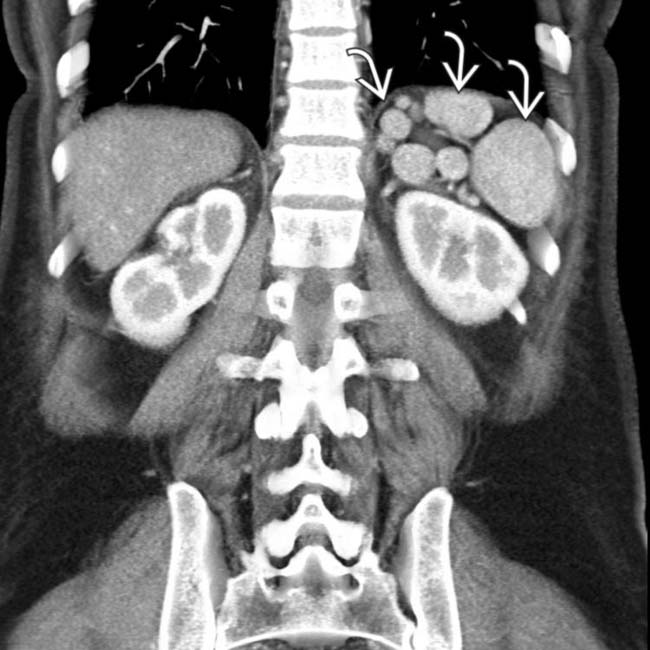
 in the left upper quadrant. The multiple spleens in PSP are typically in the left abdomen, but can rarely be on the right.
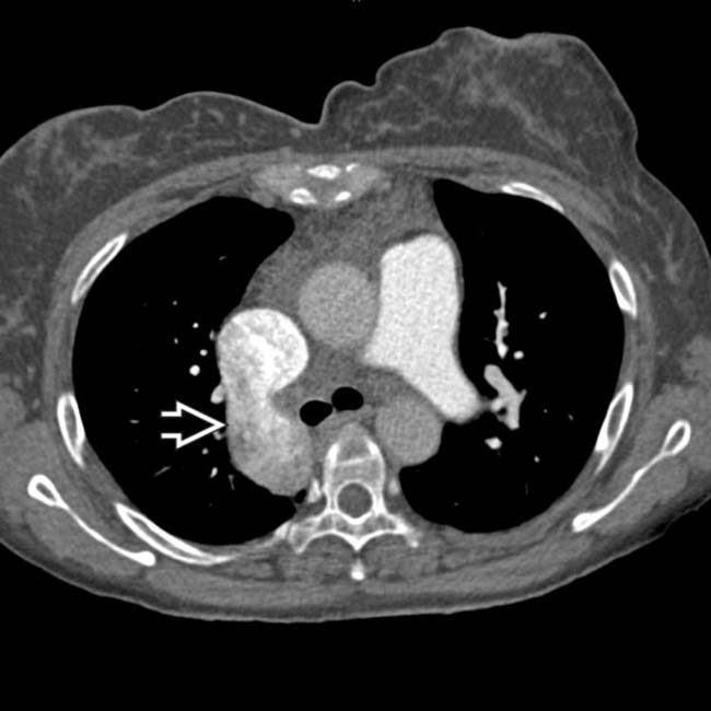
 .
.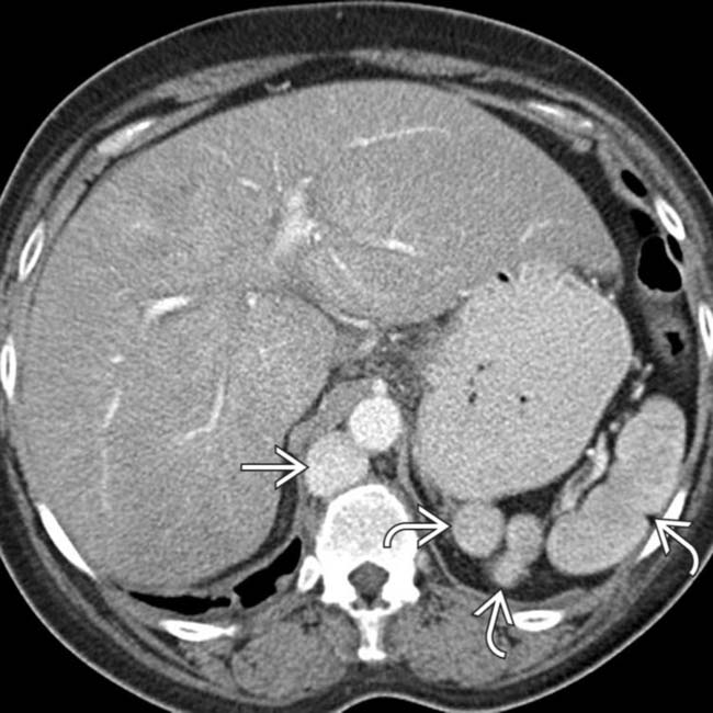
 and a dilated azygous vein
  to the right of the aorta. Azygous continuation of the inferior vena cava (IVC) is a very common abnormality in PSP syndrome.
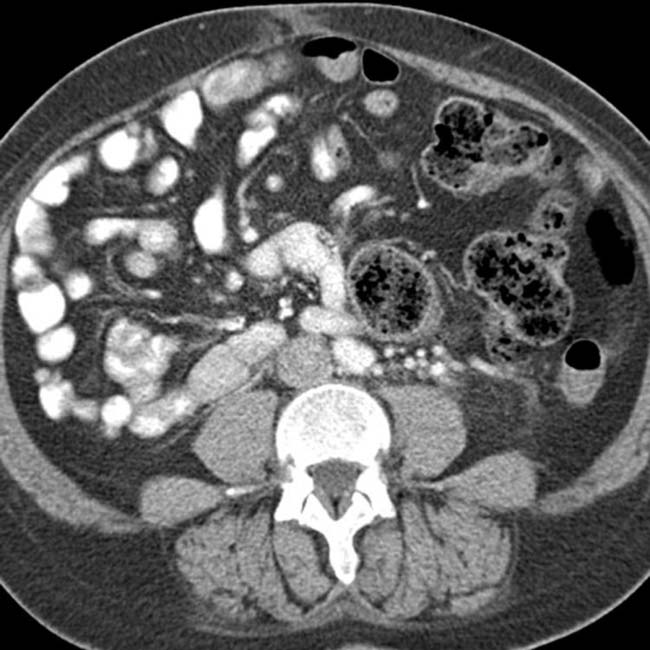


 ASP: Absence of spleen, abdominal aorta and inferior vena cava (IVC) on same side (usually right), and bilateral distribution of right-sided viscera

 ASP syndrome: Right isomerism or bilateral right-sidedness
 PSP syndrome: Left isomerism or bilateral left-sidedness

 Total anomalous pulmonary venous return (almost 100%), endocardial cushion defect (85%), single ventricle (51%), transposition of great vessels (58%), pulmonary stenosis or atresia (70%), dextrocardia (42%), mesocardia, ventricular septal defect, single atrioventricular valve, bilateral superior vena cava (SVC)


 Increased risk of complex cardiac anomalies, although less common with PSP than with ASP, accounting for better long-term survival


