CT shows diverticula as outpouchings from colonic wall, filled with gas or feces
Circular muscle hypertrophy (myochosis coli) causes irregularly spaced indentations and narrowing of lumen of colon
TOP DIFFERENTIAL DIAGNOSES
• Giant sigmoid diverticulum
Represents chronic, walled-off abscess that communicates with colonic lumen
• Diverticulitis
Due to perforation of 1 or more diverticula
PATHOLOGY
• Etiology
Sedentary lifestyle, high-fat, low-fiber diet predispose to diabetes, obesity, and diverticulosis among other ailments
CLINICAL ISSUES
• Affects > 50% of population > 60 years of age in USA
• Most common colonic disease in Western world
Diverticulosis is increasing in prevalence parallel to obesity epidemic
Diverticulosis in patients 20-40 years old is no longer rare
• Most common signs/symptoms
Most often asymptomatic
Alternating constipation and diarrhea (with circular muscle hypertrophy)
Most common cause of rectal bleeding in patients > 40 years of age
Diverticulitis or abscess
DIAGNOSTIC CHECKLIST
• Consider
Diverticulitis, if pericolonic fat stranding and pericolonic fluid are present
Colon cancer if rectal bleeding is present
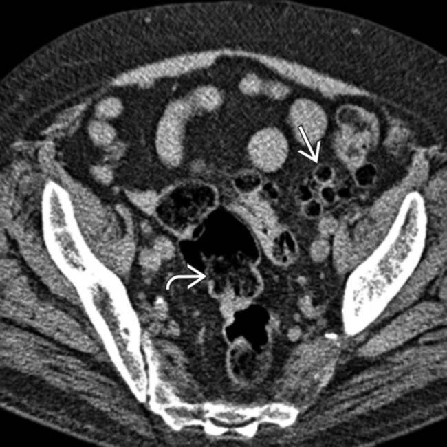
(Left) Axial NECT in a 74-year-old man with a 10-year history of obstipation presenting with lower abdominal pain shows a large amount of stool in the sigmoid colon and a cluster of gas-filled diverticula in the descending colon.
(Right) Axial CECT in the same patient illustrates the normal fat planes of the sigmoid mesocolon and the small bowel mesentery , without evidence of mural thickening or pericolonic stranding to suggest diverticulitis.
(Left) Axial CECT in a patient presenting with constipation and intermittent painful lower abdominal cramps shows thickening of the sigmoid colon wall due to myochosis, a combination of hypertrophy of the circular muscle layer, shortening of the taeniae, and lumen narrowing.
(Right) Spot film from an air-contrast BE shows distortion of the colonic lumen. The luminal outpouchings are diverticula, while the irregularly spaced infoldings of the wall represent myochosis (circular muscle hypertrophy).
TERMINOLOGY
Synonyms
• Diverticular disease
Definitions
• Outpouching of colonic mucosa and submucosa, most commonly in sigmoid colon
• Myochosis coli is uncommonly used term to describe foreshortening of colon and circular muscle hypertrophy that commonly occurs along with diverticulosis
IMAGING
General Features
• Best diagnostic clue
Rounded or oval colonic wall outpouchings
• Location
Primarily sigmoid colon, but may occur in any segment except rectum
– Lack of diverticula in rectum is due to fusion of taenia providing strong supporting coat to rectal wall
• Size
5-10 mm in diameter
• Morphology
Oval or rounded
Imaging Recommendations
• Best imaging tool
CT and barium enema (BE)
• Protocol advice
Good bowel preparation is necessary to avoid misdiagnosis of polyp vs. diverticula on air-contrast barium enema
Fluoroscopic Findings
• Contrast enema
Diverticula project out beyond wall of colon
Circular muscle hypertrophy (myochosis coli) causes irregularly spaced indentations and narrowing of lumen of colon
Easier to distinguish colonic diverticula from polyps on single contrast BE than on air-contrast BE
– Diverticula fill with barium on single contrast BE
– Diverticulum with large neck; may resemble sessile polyp on air-contrast BE
– May cause “bowler hat” sign
If “bowler hat” points to lumen, polyp likely
– Air-filled diverticula project out from bowel lumen
– Appearance of diverticula varies depending on degree of air vs. barium in diverticulum
Radiographic Findings
• Radiography
“Bubbly” appearance of sigmoid in 50% of cases
– Associated with calcified pelvic phleboliths
Diverticula arise adjacent to taenia coli
– Weakness in bowel wall due to penetration of vasa rectae
CT Findings
• Outpouchings (diverticula) filled with air, stool, or contrast agent
• Mural thickening due to myochosis (circular muscle hypertrophy) usually > 4 mm
 Circular muscle hypertrophy (myochosis coli) causes irregularly spaced indentations and narrowing of lumen of colon


 and a cluster of gas-filled diverticula
  in the descending colon.
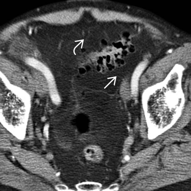
 and the small bowel mesentery
  , without evidence of mural thickening or pericolonic stranding to suggest diverticulitis.
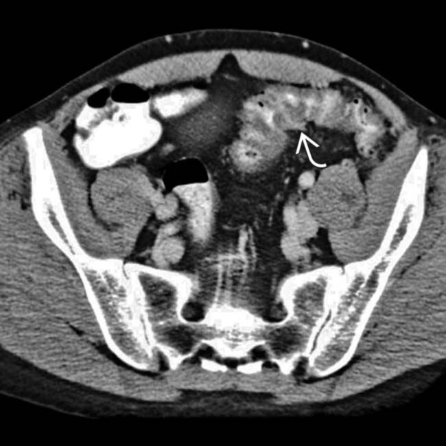
 due to myochosis, a combination of hypertrophy of the circular muscle layer, shortening of the taeniae, and lumen narrowing.
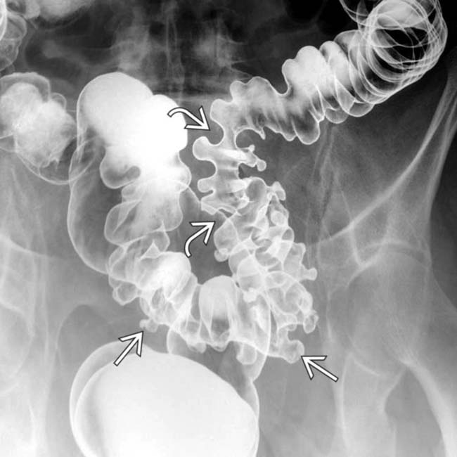
 are diverticula, while the irregularly spaced infoldings of the wall
  represent myochosis (circular muscle hypertrophy).


