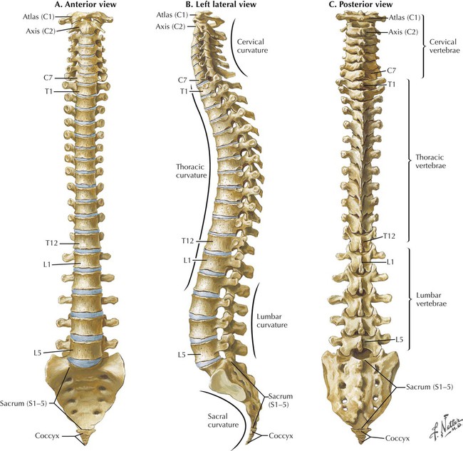
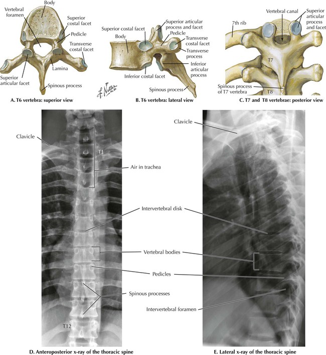
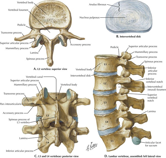
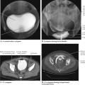
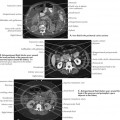
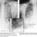
2 Back and Spinal Cord 2.1. VERTEBRAL COLUMN 2.2. THORACIC VERTEBRAE 2.3. LUMBAR VERTEBRAE 2.4. LUMBAR VERTEBRAE IMAGES 2.5. SPINAL MEMBRANES AND NERVE ORIGINS 2.6. SPINAL NERVE ORIGINS: CROSS SECTIONS 2.7. LUMBOSACRAL REGION LIGAMENTS 2.8. NERVE ROOTS 2.9. NORMAL T1 MRI STUDIES OF THE LUMBAR VERTEBRAL COLUMN 2.10. T2 AND FAT SATURATION MRI SEQUENCES 2.11. LUMBAR DISK HERNIATION 2.12. MRI OF A HERNIATED DISK 2.13. CT OF OSTEOPOROSIS IN THE THORACIC SPINE 2.14. MRI OF METASTATIC DISEASE IN THE THORACIC SPINE 2.15. MRI OF SPONDYLOLISTHESIS 2.1 Vertebral Column There are seven cervical vertebrae, twelve thoracic vertebrae defined by their articulation with the twelve pairs of ribs, five lumbar vertebrae, five fused sacral vertebrae that comprise the sacrum, and three to four fused vertebrae that form the coccyx. The cervical and lumbar vertebrae form a curve that is convex anteriorly (lordosis), whereas the thoracic vertebrae have a curve that is convex posteriorly (kyphosis). The two lordoses are secondary curves that develop postnatally. 2.2 Thoracic Vertebrae A typical vertebra consists of a body and vertebral arch enclosing a vertebral foramen that contains the spinal cord. The arch consists of pedicles and laminae, and extending from the arch are bony projections called transverse and spinous processes. Thoracic vertebrae are characterized by their facets for the articulation with ribs. The heads of ribs articulate with superior and inferior costal facets on adjacent bodies (two demifacets), and the tubercles of ribs articulate with the facets on the thick transverse processes. The thoracic spinous processes are long and slope inferiorly. The laminae are broad and flat, and the articular facets between vertebrae are oriented in a coronal plane. Lower-density, darker features in the x-rays are the intervertebral disks and the intervertebral foraminae between adjacent pedicles seen in lateral view. Pedicles appear as circular profiles in an anteroposterior view. 2.3 Lumbar Vertebrae Lumbar vertebrae have no rib articulations and are the largest vertebrae because they bear the most weight. Without costal articular facets, their transverse processes are small. Their spinous processes are horizontal in orientation and rectangular in shape. Intervertebral disks are comprised of two parts: a fibrous outer anulus fibrosus and a gelatinous inner nucleus pulposus. 2.4 Lumbar Vertebrae Images Vertebral bodies, spines, pedicles, and intervertebral foramina are evident in the x-rays. Compare the x-rays with the computed tomography (CT) sagittal reconstruction. The latter is a bone window that shows good contrast between the compact cortical bone on the surface of each vertebra and the spongy bone on the interior. Soft tissues such as muscle, intervertebral disks, the spinal cord, and cerebrospinal fluid (CSF) are not seen clearly. X-rays also have soft tissue shadowing superimposed over the bony vertebral column, which is not present in a CT digital reconstruction. Note that the plane of section in the CT is near the midline in the lumbar region but through pedicles and intervertebral foramina higher up, suggesting that there may be some scoliosis present. Abnormal bony growths (osteophytes) are seen anteriorly on the L3 and L4 lumbar vertebral bodies. Only gold members can continue reading. Log In or Register to continue Share this: Share on X (Opens in new window) X Share on Facebook (Opens in new window) Facebook Related posts: Introduction to Imaging Modalities Pelvis and Perineum Abdomen Lower Limbs Thorax Head and Neck Stay updated, free articles. Join our Telegram channel Join Tags: Netters Introduction to Imaging Jan 10, 2016 | Posted by admin in RADIOGRAPHIC ANATOMY | Comments Off on Back and Spinal Cord Full access? Get Clinical Tree
2 Back and Spinal Cord 2.1. VERTEBRAL COLUMN 2.2. THORACIC VERTEBRAE 2.3. LUMBAR VERTEBRAE 2.4. LUMBAR VERTEBRAE IMAGES 2.5. SPINAL MEMBRANES AND NERVE ORIGINS 2.6. SPINAL NERVE ORIGINS: CROSS SECTIONS 2.7. LUMBOSACRAL REGION LIGAMENTS 2.8. NERVE ROOTS 2.9. NORMAL T1 MRI STUDIES OF THE LUMBAR VERTEBRAL COLUMN 2.10. T2 AND FAT SATURATION MRI SEQUENCES 2.11. LUMBAR DISK HERNIATION 2.12. MRI OF A HERNIATED DISK 2.13. CT OF OSTEOPOROSIS IN THE THORACIC SPINE 2.14. MRI OF METASTATIC DISEASE IN THE THORACIC SPINE 2.15. MRI OF SPONDYLOLISTHESIS 2.1 Vertebral Column There are seven cervical vertebrae, twelve thoracic vertebrae defined by their articulation with the twelve pairs of ribs, five lumbar vertebrae, five fused sacral vertebrae that comprise the sacrum, and three to four fused vertebrae that form the coccyx. The cervical and lumbar vertebrae form a curve that is convex anteriorly (lordosis), whereas the thoracic vertebrae have a curve that is convex posteriorly (kyphosis). The two lordoses are secondary curves that develop postnatally. 2.2 Thoracic Vertebrae A typical vertebra consists of a body and vertebral arch enclosing a vertebral foramen that contains the spinal cord. The arch consists of pedicles and laminae, and extending from the arch are bony projections called transverse and spinous processes. Thoracic vertebrae are characterized by their facets for the articulation with ribs. The heads of ribs articulate with superior and inferior costal facets on adjacent bodies (two demifacets), and the tubercles of ribs articulate with the facets on the thick transverse processes. The thoracic spinous processes are long and slope inferiorly. The laminae are broad and flat, and the articular facets between vertebrae are oriented in a coronal plane. Lower-density, darker features in the x-rays are the intervertebral disks and the intervertebral foraminae between adjacent pedicles seen in lateral view. Pedicles appear as circular profiles in an anteroposterior view. 2.3 Lumbar Vertebrae Lumbar vertebrae have no rib articulations and are the largest vertebrae because they bear the most weight. Without costal articular facets, their transverse processes are small. Their spinous processes are horizontal in orientation and rectangular in shape. Intervertebral disks are comprised of two parts: a fibrous outer anulus fibrosus and a gelatinous inner nucleus pulposus. 2.4 Lumbar Vertebrae Images Vertebral bodies, spines, pedicles, and intervertebral foramina are evident in the x-rays. Compare the x-rays with the computed tomography (CT) sagittal reconstruction. The latter is a bone window that shows good contrast between the compact cortical bone on the surface of each vertebra and the spongy bone on the interior. Soft tissues such as muscle, intervertebral disks, the spinal cord, and cerebrospinal fluid (CSF) are not seen clearly. X-rays also have soft tissue shadowing superimposed over the bony vertebral column, which is not present in a CT digital reconstruction. Note that the plane of section in the CT is near the midline in the lumbar region but through pedicles and intervertebral foramina higher up, suggesting that there may be some scoliosis present. Abnormal bony growths (osteophytes) are seen anteriorly on the L3 and L4 lumbar vertebral bodies. Only gold members can continue reading. Log In or Register to continue Share this: Share on X (Opens in new window) X Share on Facebook (Opens in new window) Facebook Related posts: Introduction to Imaging Modalities Pelvis and Perineum Abdomen Lower Limbs Thorax Head and Neck Stay updated, free articles. Join our Telegram channel Join Tags: Netters Introduction to Imaging Jan 10, 2016 | Posted by admin in RADIOGRAPHIC ANATOMY | Comments Off on Back and Spinal Cord Full access? Get Clinical Tree