Fig. 1.1
Figure displaying normal ostia of the left and right coronary arteries arising from the left and right coronary cusps, respectively. Notice the ostia arise between the margin of the aortic valve leaflets and sinotubular junction. The coronary arteries include: R right coronary, L left coronary, LM left main, LAD Left anterior descending. The aortic valve cusps: R right, L left, NC noncoronary (or posterior) (From Waller et al. [8] with permission)
Coronary ostia typically arise from the middle of the right and left sinus of valsalva; below the sinotubular junction and above the free margin of the corresponding open aortic valve leaflet [1, 8]. This allows for maximal coronary filling during diastole. A coronary ostium that that arises above or below the sinus of valsalva is termed to be a variant of normal anatomy (Fig. 1.2). If the ostium of a coronary artery takes off >1 cm above the sinotubuluar junction, it is considered a high take off or ectopic position [9]. This has been described to be associated with decreased diastolic filling and chronic ischemia in the absence of epicardial stenosis [10].
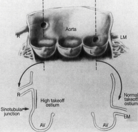

Fig. 1.2
Figure displays the normal take off of the left main and high takeoff off of the right coronary artery. Each artery arises from the proper coronary cusp-the right and left coronary arteries arise from the right and left coronary cusps, respectively. R right, L left, LM left main, AV aortic valve (From Waller et al. [8] with permission)
Normally, there are two to three coronary ostia [11]. Two ostia are more common and correspond with the left and right coronary arteries. The third typically comes from a separate ostium for the conus or infundibular branch that is present in 23–51 % or normal hearts [1, 12] and has been referred to as the “third coronary artery”. Less commonly, there is an absence of the left main with separate ostia of the left anterior descending and the left circumflex arteries (Fig. 1.3). The ostial orientation is generally orthogonal to the aortic root or ascending aorta [6]. Although there is some variation, the right coronary artery ostium generally arises in the vertical plane and the left coronary in the horizontal plane (Figs. 1.4, 1.5, and 1.6).
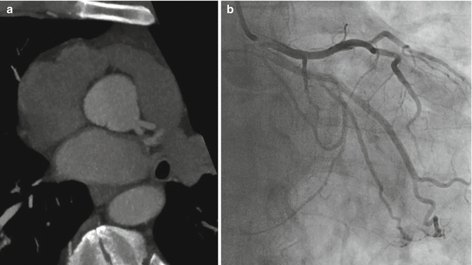
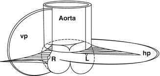
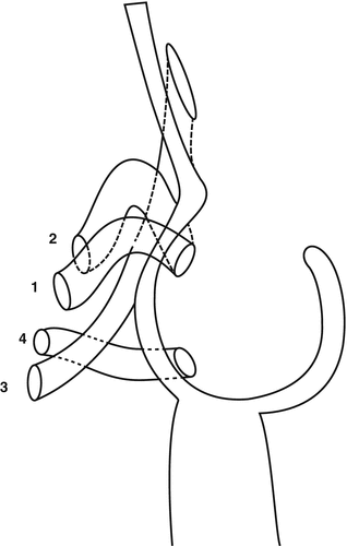
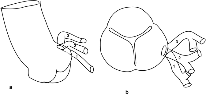

Fig. 1.3
Figures demonstrate an absent left main with the left anterior descending and left circumflex arteries arising from separate ostia in the left sinus of valsalva (a) left coronary CTA and (b) right selective coronary angiography

Fig. 1.4
This figure represents the coronary orientation in regard to the aortic root and ascending aorta. The right and left coronary artery ostia are oriented in a vertical plane (vp) and horizontal plane (hp), respectively (From Angelini [6] with permission)

Fig. 1.5
This represents a cross sectional view of the variable right coronary ostium orientation. (1) Normal and remains orthogonal to the aorta in the vertical plane (2) Upward takeoff (3) Downward takeoff (4) Horizontal orientation (From Angelini [6] with permission)

Fig. 1.6
The orientation of the left coronary artery ostium in the frontal (a) and horizontal (b) planes. (1) Inferior tilt (2) Normal orthogonal orientation (3) Superior tilt (From Angelini [6] with permission)
Myocardial Bridging
Normal epicardial coronary arteries occasionally take a short intramyocardial course. This causes arterial compression during systole referred to as milking or systolic “myocardial bridging”. Although this can occur in any vessel, it is most commonly seen in the LAD. Myocaridal bridging is reported as frequently as 25 % by autopsy studies and 2 % angiographically [13–15]. Generally, myocardial bridging is considered a benign phenomenon, as the 5-year survival remains high with rare reports of sudden cardiac death. Despite the fact that much of the coronary compression occurs during systole and the majority of coronary perfusion occurs during diastole, there are reports of underlying ischemia driven by myocardial bridging [16, 17]. This has been described in patients with long segments of an intramyocardial course. Increased heart rates and decreased diastolic filling pressures contribute to ischemia by decreasing diastolic filling time and increased systolic coronary compression, respectively.
The Coronary Arteries
Right Coronary Artery
The right coronary artery (RCA) arises anteriorly from the right coronary cusp and travels anteriorly and posteriorly in the atrioventricular groove [18, 19] (Figs. 1.7 and 1.8). If the RCA is the dominant vessel, it travels posteriorly and provides branches along the interventricular groove and lateral wall of the left ventricle; the posterior descending artery and posterolateral branch, respectively.
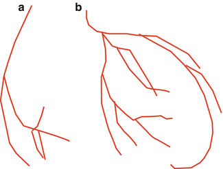
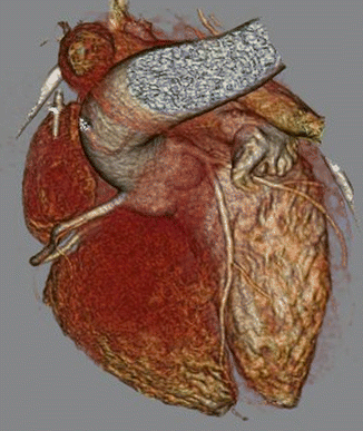

Fig. 1.7
Wire model of coronaries (a) RCA in LAO projection; (b) the Left coronary system in the RAO projection

Fig. 1.8
This figure demonstrates normal coronary anatomy. The left and right coronary arteries arise from the respective aortic cusps. The left anterior descending artery courses anteriorly between the left and right ventricles. The left circumflex and right coronary arteries travel in the left and right atrioventricular grooves, respectively. LAD left anterior descending, RCA right coronary artery
The usual dominant RCA is 12–14 cm in length prior to giving off a PDA [20]. The luminal diameter generally ranges from 1.5 to 5.5 mm with a mean of 3.2 mm [20]. While the LAD and LCX tend to taper as they progress distally, the diameter of the RCA remains relatively constant until just prior to the take off of the PDA. The first branch of the RCA is the infundibular or conus branch in 50 % of the population. This supplies the right ventricular outflow tract and often anastomoses with an infundibular branch of the left anterior descending artery forming the circle of Vieussens [21]. In the other half, the conus branch arises from a separate ostium in the right coronary sinus of Valsalva. In 60 % of the population, the second branch of the RCA is the sinus nodal artery [22]. In the remaining 40 %, the sinus nodal artery is a branch from the circumflex artery. The RCA then gives off small branches supplying the right atrium and ventricle. The largest of these is the acute marginal artery; which supplies much of right ventricular free wall [23]. If the RCA is dominant, it supplies two several major branches: (1) the posterior descending artery (2) posterolateral branch. The posterior descending artery travels in the posterior interventricular grove and supplies the posterior inferior septum. If the left anterior descending artery does not reach the apex of the heart, the PDA can supply the distal third of the interventricular septum. The posterolateral branch (es) supply the lateral wall. Just distal to the PDA, the RCA occasionally supplies an AV nodal branch [8].
Left Main Artery
The left main (LM) artery originates from the left sinus of Valsalva and travels anteriorly and leftward (Figs. 1.7 and 1.8). It is positioned between the left atrial appendage and the pulmonary trunk [5]. This divides into two major branches: the left anterior descending (LAD) and left circumflex (LCX) arteries. The LM varies in length from 0.5 to 2.5 cm but remains uniform in caliber throughout its length [20, 24, 25]. The LM can trifurcate providing a third branch referred to as a ramus intermedius (RI) (Fig. 1.9). The RI originates between the LAD and the LCX and supplies the territory of the obtuse marginal and/or the diagonal [25]. The luminal diameter of the LM is usually 2.0–5.5 mm with a mean of 4 mm [20].
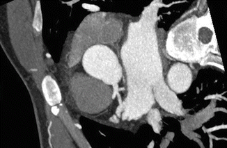

Fig. 1.9
Ramus Intermedius. In some individuals, instead of the typical bifurcation into the LAD and LCX, the left main trifurcates into an LAD, LCX, and ramus intermedius (RI), the RI coursing between the LAD and LCX. This can be difficult at times to differentiate from a early diagonal branch of the LAD or obtuse marginal of the LCX
Left Anterior Descending Artery
The LAD extends from the left main and curves around the pulmonary trunk prior to entering the anterior interventricular groove and extending to the apex [26]. The left anterior descending artery then extends distally to the apex within the inferior interventricular sulcus towards the crux of the heart. It then provides branches to the inferior walls of both ventricles [26]. The vessel terminates in the interventricular groove prior to the posterior (inferior) descending artery (Figs. 1.7 and 1.8). The anterior descending artery provides two major branches: septal perforator arteries and diagonal branches. The septal perforator arteries branch at right angles from the anterior descending artery and supply the anterior two thirds of the intraventricular septum [22]. The diagonal branches are typically larger than the septal perforators and supply the lateral wall of the left ventricle. The diagonal branches are sequentially numbered as they arise from the LAD. The anterior descending artery can also produce an infundibular/conal branch. The LAD is generally has a luminal area from 2.0 to 5.0 mm with an average of 3.6 mm [20].
Left Circumflex Artery
The left circumflex artery has a branching angle from the main stem that is variable. It then courses through the left atrioventricular groove [26]. This artery provides obtuse marginal branches that are sequentially numbered as they arise from the LCX and supply the posterior and lateral wall of the left ventricle (Figs. 1.7 and 1.8). If this is the dominant vessel, it provides the PDA and PLB; rather than the right coronary artery (Fig. 1.10). The luminal area of the LCX is generally 1.5–5.5 mm with an average of 3.2 mm [20].
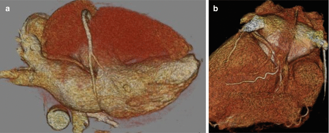

Fig. 1.10
Coronary Dominance. Coronary dominance is determined by the vessel that supplies the posterior descending (PDA) and posterolateral branches (PLB) to the inferior wall of the left ventricle. The right coronary artery is considered dominant if it supplies both the PDA and PLB as seen in (a). The left circumflex is considered dominant if it supplies both as seen in (b). It is considered a co-dominant system if the RCA supplies the PDA and the LCX supplies the PLB
Dominance
The vessel that supplies the PDA and PLB determines coronary dominance (Fig. 1.10). The artery that supplies both vessels is considered the dominant vessel. If one provides the PDA and the other provides the PLB, it is considered to be co-dominant or have balanced dominance. In the general population, dominance of the right coronary artery is most common and has been described in up to 89 % of the population. The left coronary artery is dominant in approximately 7–8 % of the population [27–30]. A co-dominant system was noted in approximately 4 % of the population. The clinical significance of dominance is not entirely clear, though there have been data suggesting increased adverse events (cardiovascular related mortality and non-fatal MI) with those who are left dominant [30]. Some data suggest increased incidence of perfusion defects on nuclear studies.
Stay updated, free articles. Join our Telegram channel

Full access? Get Clinical Tree


