Case presentation
A 14-month-old female is referred from her primary care pediatrician for increasing abdominal girth and an abdominal mass. For the past month, the child’s abdomen has been increasing in size, as noted by her parents. There has been no associated fever, vomiting, diarrhea, trauma, back pain, urinary complaints, difficulty breathing, or cough. She is otherwise behaving at her baseline.
Physical examination reveals a well-appearing child in no distress. Her vital signs are unremarkable and appropriate for her age. She has a remarkable abdominal examination: There is obvious abdominal distention and a palpable right flank mass. There is no tenderness, guarding, or rebound.
Imaging considerations
Ultrasound (US)
This imaging modality is a first-line imaging choice in pediatric patients suspected of having an intraabdominal mass. , The study is generally readily available and requires no sedation to complete. Additionally, there is no exposure to ionizing radiation and US can differentiate between abdominal masses and other causes of mass effect such as hydronephrosis, distended bowel, or a distended urinary bladder. These attributes make ultrasonography an excellent screening modality. Wilms tumor usually appears as a well-defined mass arising from the kidney; however, when the mass is very large, the site of origin can be difficult to determine. Doppler flow studies should also be obtained at the time of imaging, since Wilms tumor can invade the renal vein, the inferior vena cava, or, rarely, the right atrium (termed intracardiac Wilms tumor).
Plain radiography
Plain radiography may initially be used in patients with abdominal distention. Findings may be nonspecific and masses may or may not be visualized. As a general rule, plain radiography of the abdomen is not useful for abdominal mass detection per se but will provide a view of the bowel gas pattern, may detect calcifications, and may demonstrate mass effect if an intraabdominal tumor is present, prompting additional imaging with US or computed tomography (CT).
If an abdominal mass suspicious for Wilms tumor is found on imaging, chest x-ray may be utilized as the lungs are a primary site of metastasis for this tumor.
Computed tomography
CT is an excellent imaging modality used to evaluate intraabdominal pathology. While readily available and rapid, the patient is exposed to ionizing radiation. Intravenous contrast should be utilized. CT of the abdomen and pelvis should be obtained if ultrasonography demonstrates a renal mass; if Wilms tumor is suspected, additional CT of the chest may be obtained, since the lung is a common site of metastases for this lesion, with up to 15% of patients having lung involvement at the time of diagnosis. CT of the chest may also be utilized if plain chest radiography is abnormal. , , Wilms tumor is visualized as a solid mass arising from the kidney, with renal parenchyma splayed around the border with the mass (the “claw sign”); calcifications within the tumor are possible, but not commonly seen. Once treatment is completed, CT is used to monitor for recurrence.
The decision to utilize plain radiography or CT for chest imaging may be determined after consultation with Pediatric Oncology and/or Pediatric Surgery .
Magnetic resonance imaging (MRI)
This imaging modality, like CT, is utilized as a secondary test when US is abnormal. Like CT, MRI provides visualization of the contralateral kidney and intraabdominal contents. However, availability may be a barrier and sedation is generally required to complete a meaningful study. MRI is often utilized as a surveillance imaging modality.
Imaging findings
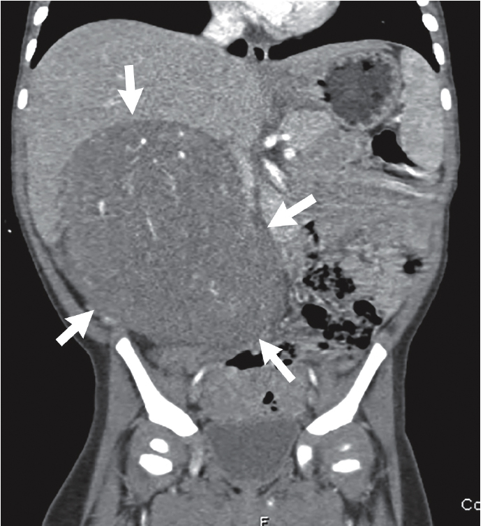
The CT study (with intravenous contrast) obtained by the primary care provider was reviewed. Selected images are provided here. There is a large renal mass, which is solid in appearance; the remaining intraabdominal organs appear normal ( Figs. 25.1–25.3 ).