George M. Bridgeforth and Mark Nolden
AN 18-year-old man is involved in a motor vehicle collision in which he is rear-ended. he is disoriented and complains of severe cervical pain as well as weakness and numbness of the arms and legs.
CLINICAL POINTS
- This condition is considered unstable.
- Associated neurological deficits are usually present; cervical injuries may cause quadriplegia.
- Disk herniation may occur.
Clinical Presentation
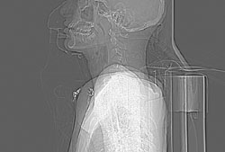
Bilateral facet dislocations are unstable injuries characterized by disruption of the posterior ligament complex and damage to the anterior and posterior longitudinal ligaments. The cervical canal is no longer intact. The mechanism of injury is hyperflexion. Most of the injuries affect the midcervical region. Thoracic and lumbar injuries are rare but have been reported. As a result of the soft tissue damage, neurological injury is common.
Within 48 to 72 hours, the acute spinal shock is followed by upper motor neuron signs of motor (deltoid) weakness, hyperreflexia, clonus (oscillating reflexes), spasticity, and upgoing plantar responses (upgoing big toe when the soles of the feet are stroked with a reflex hammer). High-level spinal cord injuries (C2–C4) are characterized by damage to the phrenic nerve and severe permanent respiratory impairments. Patients with high-level injuries are ventilator-dependent tetraplegics (quadriplegics). Unlike paraplegics, tetraplegics cannot live independently. Even after extensive training, they require assistance with daily living.
Radiographic Evaluation
Tests to order, for both acute cervical trauma and acute thoracolumbar trauma include anteroposterior, lateral, and oblique views (two). In cases of cervical injury, it is necessary to see to C7. Optional radiographs include the following:
- Swimmer view, which is used to visualize the disc space between C-7 and T-1 (C7) if it is not identified on the lateral projection
- Flexion/extension views, which should be obtained only by experienced spinal care providers. Flexion and extension radiographs are never taken until the initial radiographs have been evaluated first for instability.
PATIENT ASSESSMENT 
- With acute injuries, possible weakness, sensory loss, loss of sphincter tone, and loss of reflexes
- Associated head injuries and organ damage
- Progressive paralysis
Related posts:


Stay updated, free articles. Join our Telegram channel

Full access? Get Clinical Tree


