George M. Bridgeforth and George Holmes
A 55-year-old woman slipped and fell on her icy driveway yesterday. She presents with pain over the base of the left fifth metatarsal.
CLINICAL POINTS
- Patients complain of pain and swelling.
- Weight-bearing is limited.
- Tenderness at the base of the fifth metatarsal may be present.
Clinical Presentation
Fractures of the foot are common and often involve the fifth metatarsal, the bone that runs from the middle of the foot to the base of the small toe (Fig. 25.1). There are basically three types of fifth metatarsal injuries.
- Avulsion fractures: These fractures have a mechanism of injury that is similar to an ankle sprain.* Patients may complain of tripping or missing a curb or the rung of ladder. Avulsion fractures are transverse fractures that generally involve the tuberosity of the metatarsal base—the site of attachment of the avulsed peroneus brevis tendon.
- Jones fractures: True Jones fractures, more rare than avulsion fractures, occur at the proximal diaphysis (shaft). These result from laterally directed force on the forefoot with the ankle in plantar flexion (see “Radiographic Evaluation” for more information); they are caused by inversion of the foot. The resulting injury is more serious (Fig. 25.2).
- Stress fractures: Stress fractures of the metatarsals may occur distally at the metatarsal neck in runners. However, some stress fractures may appear more proximally, especially in dancers; axial loads with torsion result in more proximal fractures. These result from the placement of abnormal stress on a normal bone.
PATIENT ASSESSMENT 
- Swelling and point tenderness with an inability to bear weight along the lateral border of the foot
- Sometimes caused by placement of abnormal stress
- Easily missed in many cases, because the examiner focuses on the ankle injury. The examiner should always check for point tenderness of the metatarsal tenderness with any ankle injury.
Patients with a fifth metatarsal styloid fracture or a Jones fracture (proximal diaphysis) exhibit marked tenderness at the proximal fifth metatarsal, and bruising may be present. In the case of fifth metatarsal styloid fracture, the pain and soreness is at the tuberosity of the fifth metatarsal base. There is pain-limited weight-bearing along the lateral aspect of the foot.
When examining a patient for a fracture of the fifth metatarsal, the examiner should
- specifically palpate for a fifth metatarsal fracture. An associated metatarsal fracture is easily missed because the examiner focuses on the pain and swelling caused by the ankle injury.
- observe the Ottawa rules. Look for pain, swelling, or tenderness of the lateral malleolus or both malleoli. It is important to check for navicular and fibular head tenderness as well.
- always check and record the neurovascular status
Radiographic Evaluation
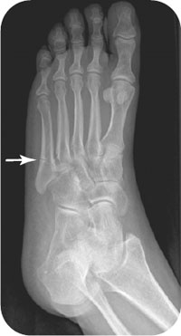
Radiographs to order include anteroposterior (AP), lateral, and oblique views. If there is associated metatarsal tenderness of the base or proximal shaft and the area is not well visualized on standard ankle radiographs, then AP, lateral, and oblique foot radiographs should be obtained as well. It is always necessary to check all foot radiographs (and lateral ankle radiographs) for a fifth metatarsal fracture (Fig. 25.3). An avulsion fracture usually is located at or proximal to the metatarsal–cuboid joint (Fig. 25.4). True Jones fractures must be distinguished from avulsion fractures at the base of the fifth metatarsal. Also, true Jones fractures are rare and occur near (about 1 inch distal to) the metaphyseal–diaphyseal junction (i.e., right at the proximal fifth metatarsal shaft) (Fig. 25.5). This region must be visualized on all ankle radiographs. Generally, Jones fractures are at or distal to a transverse line drawn from the metatarsal–cuboid joint. The zonal classification is helpful in this case. Fractures at the tuberosity of the fifth metatarsal are avulsion fractures (zone 1). Fractures at the proximal diaphysis (shaft) are Jones fractures (zone 2). Fractures of the mid or distal shaft are stress fractures (zone 3) (Fig. 25.6).
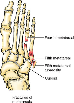
FIGURE 25.1 Anatomical diagram demonstrating a displaced transverse of the fourth metatarsal shaft. Note the avulsion fracture of the fifth metatarsal tuberosity. Jones fractures are uncommon but are characterized by transverse fractures of the proximal shaft (diaphysis) of the fifth metatarsal. (From Moore KL, Agur A. Essential Clinical Anatomy. 2nd ed. Philadelphia, PA: Lippincott Williams & Wilkins; 2002.)
Related posts:


Stay updated, free articles. Join our Telegram channel

Full access? Get Clinical Tree


