George M. Bridgeforth, Thomas Hearty, and Charles Carroll IV
A 40-year-old man injured himself in a moped accident, where he fell onto his outstretched hand. he now presents with pain and swelling of the wrist.
CLINICAL POINTS
- Injury often involves a fall onto an outstretched hand.
- The scaphoid bone is located on the thumb side of the wrist.
- Untreated scaphoid fractures may lead to instability, premature degenerative arthritis, and necrosis of the proximal scaphoid.
Clinical Presentation
As stated in the previous chapter, scaphoid fractures are the second most common fracture to the distal forearm/wrist (behind radial fractures). Fractures of the carpal bones account for approximately 6% of all fractures, and the scaphoid is the carpal bone fractured most often. Scaphoid fractures are most likely to occur in men who are 20 to 30 years of age. Like Colles fractures, scaphoid (navicular) fractures are usually caused by a fall onto an outstretched hand with the wrist in hyperextension, but they can result in other ways.
Pain, swelling, and tenderness (as well as bruising when present) are characteristic signs that point the examiner to the fracture. The combination of snuffbox tenderness, scaphoid tubercle tenderness, and pain with axial compression is 100% sensitive for a scaphoid fracture.
Note that patients with both Colles fractures and scaphoid fractures may present with pain with limited range of motion. However, in the case of scaphoid fractures, the pain-limited range of motion may be out of proportion to the injury unless the examiner specifically palpates for scaphoid tenderness at the anatomic snuffbox and proximal radial surface of the wrist. It is also necessary to check neurovascular status (cold cyanotic fingers, diminished or absent pulses, delayed capillary refill of over 4 seconds in fingertips) carefully. In addition, another key clinical clue helps differentiate a scaphoid fracture from a Colles fracture. Patients with Colles fractures usually have a characteristic dorsal deformity of the distal segment of the radius, whereas patients with scaphoid fractures do not.
PATIENT ASSESSMENT 
- Pain and swelling of the wrist
- Tenderness of the scaphoid
- Anatomic snuffbox (inaccurate finding if in isolation)
- Scaphoid tubercle
- Anatomic snuffbox (inaccurate finding if in isolation)
- Pain with axial compression
- Pain-limited range of motion
Other injuries can mimic a scaphoid fracture. It is always important to check the distal radius for a small radial styloid fracture or a nondisplaced distal radial fracture (which appears as a small dark line which extends to the cortex) (see section, “Radiographic Evaluation”). Patients who complain of constant clicking with wrist movement may have a subluxating scaphoid or lunate. Those with persistent pain and soreness of the anatomic snuffbox (in the absence of a fracture/dislocation) should be evaluated and treated for de Quervain’s tendonitis. This is a tendonitis affecting the extensor and abductor tendons of the thumb. De Quervain’s tendonitis may be seen with overuse injuries or following acute trauma.
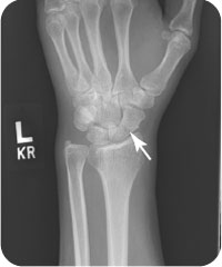
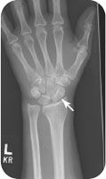
FIGURE 38.1 An oblique radiograph of the left hand of the patient in the introductory case demonstrating an acute fracture through the scaphoid.
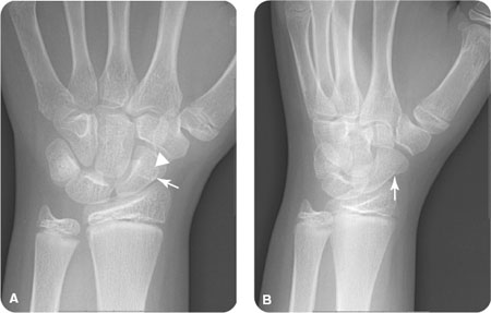
FIGURE 38.2 (A) An AP radiograph of the left wrist demonstrates a subtle overhanging edge (arrow) and dense overlapping fracture line (arrowhead) along the ulnar aspect of the scaphoid. (B) An oblique radiograph of the same patient, which better demonstrates the acute scaphoid fracture without distraction (arrow).
Related posts:


Stay updated, free articles. Join our Telegram channel

Full access? Get Clinical Tree


