George M. Bridgeforth and John Cherf
A 20-year-old intoxicated woman complains of marked pain, swelling, and tenderness after her left knee strikes the dashboard in a motor vehicle accident. She has trouble flexing this knee and complains of marked pain on standing and walking.
CLINICAL POINTS
- High-impact injuries are the usual cause of these fractures.
- With a large effusion, guarding and apprehension, which may make stability tests difficult to perform, are common.
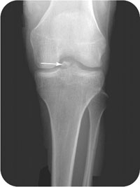
- Associated internal derangement, especially tears of the anterior or posterior cruciate ligaments, may occur (Fig. 16.1).
Clinical Presentation
Tibial spine (intercondylar) fracture is a fracture of the median eminence, which is one of two bony moundlike elevations at the proximal surface of the tibia. Structural damage to the tibial spine is commonly associated with cruciate ligament injuries. These fractures are generally caused by an anterior-to-posterior, high-impact, forces that are directed against a flexed knee, such as motor vehicle accidents and contact sports injuries. Symptoms include pain-limited range of motion, with an inability to flex or extend the knee, and reluctance to bear weight. Marked swelling of the affected knee is also evident.
Like all traumatic knee injuries it is essential that a thorough neurovascular examination of the extremity be conducted. The extremity should be examined carefully for pallor, although this feature may not be readily apparent in dark-skinned individuals. However, if the patient is coherent, he or she may complain of coldness or numbness. In addition, it is essential to check the popliteal, dorsalis pedis, and posterior tibial pulses. The popliteal pulse may be checked by placing the four fingers of each hand and palpating firmly at the center of popliteal fossa (i.e., back of the knee). The dorsal pedis pulse is located with four fingers by palpation along the medial border of the extensor hallucis longus tendon in the forefoot. The posterior tibial pulses are located by placing four fingers behind the medial malleolus near its base.
Marked swelling of the knee or lower extremity, coupled with diminished (or absent) pulses, diffuse motor weakness, and sensory impairment represents either severe neurovascular damage or secondary damage from a compartment syndrome. With a compartment syndrome, there is markedly elevated pressure and swelling inside the lower extremity, which impairs the vascular supply to the muscles and the nerves. Untreated, it can cause permanent neurological damage and, in severe cases, result in amputation of the extremity.
PATIENT ASSESSMENT 
- Pain, with limited range of motion
- Marked tenderness
- Swelling
- Antalgic gait (limp)
Septic joints that are very warm, erythematous (red), and tender do not usually follow acute traumatic injuries. However, they may be caused by open wounds in patients seen for the first time 1 or 2 days after the injury. Septic joints, with or without open wounds, should undergo an immediate diagnostic aspiration.
FIGURE 16.1 Avulsion fracture of the tibial spine in a 9-year-old girl. A significant hemarthrosis was present and was aspirated. In an adult, the same mechanism of injury would have resulted in a tear of the anterior cruciate ligament. (From Fleisher GR. Textbook of Pediatric Emergency Medicine. 5th ed. Philadelphia, PA: Lippincott Williams & Wilkins; 2005.)
Related posts:


Stay updated, free articles. Join our Telegram channel

Full access? Get Clinical Tree


