George M. Bridgeforth and George Holmes
A 35-year-old man injures his right ankle when he trips at a curb. He complains of moderate pain, swelling, and tenderness along the outside of the ankle.

CLINICAL POINTS
- Discovery of accessory ossification is incidental.
- No treatment is necessary.
Clinical Presentation
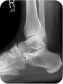
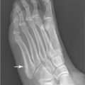
Accessory ossification centers are nontraumatic occurrences; the mechanism of injury is failure to fuse during development. They are incidental findings seen on routine radiographs. Although accessory ossification centers may appear in one of several different locations in the hindfoot and midfoot (even the forefoot), they usually occur alongside the inferior lateral border of the cuboid, the proximal tuberosity of the fifth metacarpal, or the posterior talus (Fig. 24.1). Usually, they are asymptomatic. The absence of pain, swelling, and tenderness along with their characteristic sesamoid appearance should prevent the examiner from confusing them with an acute fracture. Accessory ossification centers rarely require surgical removal.
Radiographic Evaluation
Accessory ossification centers are visible on radiographs taken for other purposes. Unlike acute fractures, they are not associated with soft swelling and tenderness and usually do not fit into larger bones like pieces of a puzzle. Common ossicles include the os peroneum (peroneal ossicle), which lies inferior laterally to the cuboid (Fig. 24.2) and the os vesalianum (vesalian ossicle), which is proximal inferior border of the tuberosity of the fifth metatarsal (Fig. 24.3). The os trigonum (trigone ossicle) lies posterior to the talus (Fig. 24.4). The peroneal ossicle and the vesalian ossicle may be identified on the lateral or the oblique view. The trigone ossicle is best appreciated on the lateral view.
PATIENT ASSESSMENT 
- Asymptomatic
- Seldom require surgery
Stay updated, free articles. Join our Telegram channel

Full access? Get Clinical Tree


