• Multiple, near water density/intensity liver lesions < 15 mm in diameter
Varied enhancement based on cystic and solid components
No communication with biliary tree
• US: Small and well-circumscribed lesions
Often have echogenic walls with small fluid content
• US shows much more echogenicity and fewer cystic lesions than anticipated based on prior CT or MR
TOP DIFFERENTIAL DIAGNOSES
• Autosomal dominant polycystic liver disease
Larger, more numerous cysts in liver and other organs
• Multiple simple hepatic cysts
Fewer cysts of varying size; no mural nodules
• Caroli disease
Central dot sign on CECT and MR
ERCP and MRCP: Communicating bile duct abnormality
• Multiple/solitary, small metastatic lesions
More complex and varied in size
• Opportunistic infection (microabscesses)
Must be considered in immunosuppressed patient with fever
DIAGNOSTIC CHECKLIST
• No further evaluation needed when seen as isolated finding in healthy, nononcologic patient
• Should be considered as likely diagnosis in setting of innumerable small, slightly complex “cysts” in healthy patient
• Lesions appear more echogenic than expected on sonography
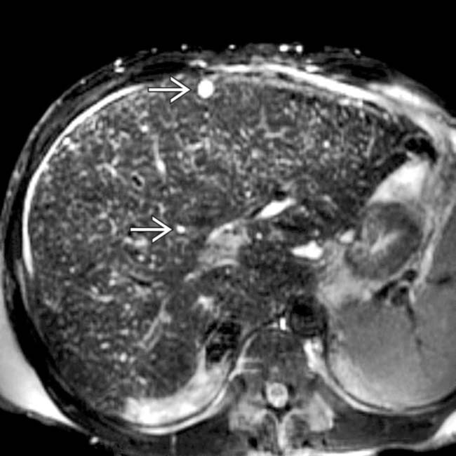
(Left) Axial T2WI MR shows innumerable tiny bright foci throughout the liver , representing biliary hamartomas. This patient also had evidence of congenital hepatic fibrosis on imaging and liver biopsy, both part of the congenital hepatic and renal fibropolycystic disease spectrum.
(Right) MRCP shows small spherical cyst-like lesions that do not communicate with the (normal) biliary tree. This feature helps to distinguish biliary hamartomas from Caroli disease.
(Left) Sonographic image shows innumerable tiny echogenic foci throughout the liver and 1 of ∼ 10 cyst-like lesions , though even these have small foci of echogenicity within the wall. MR on this patient showed many more cystic-appearing biliary hamartomas.
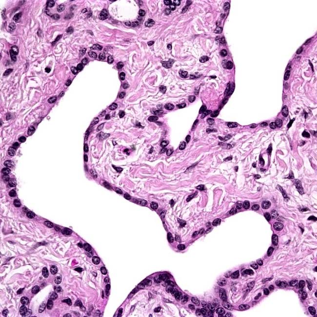
(Right) The branching, angulated glands in biliary hamartomas are lined by a single layer of flattened cuboidal epithelium. These glands may expand or rupture to produce small “cysts.” There is no nuclear atypia. (Courtesy S. Kakar, MD.)
TERMINOLOGY
Synonyms
• Bile duct microhamartoma
• von Meyenburg complex
Definitions
• Uncommon benign malformations of biliary tract
IMAGING
General Features
• Best diagnostic clue
Multiple near water density/intensity liver lesions < 15 mm in diameter
• Location
Subcapsular or intraparenchymal in location
Scattered throughout both lobes of liver
• Size
• Irregular spherical
• Usually multiple to innumerable
CT Findings
• NECT
Density of lesions depends on predominance of cystic or solid component


 , representing biliary hamartomas. This patient also had evidence of congenital hepatic fibrosis on imaging and liver biopsy, both part of the congenital hepatic and renal fibropolycystic disease spectrum.
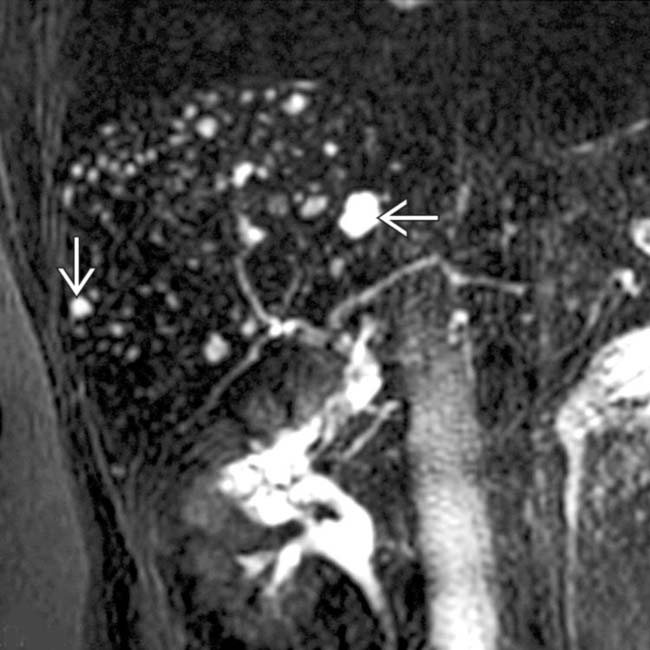
 that do not communicate with the (normal) biliary tree. This feature helps to distinguish biliary hamartomas from Caroli disease.
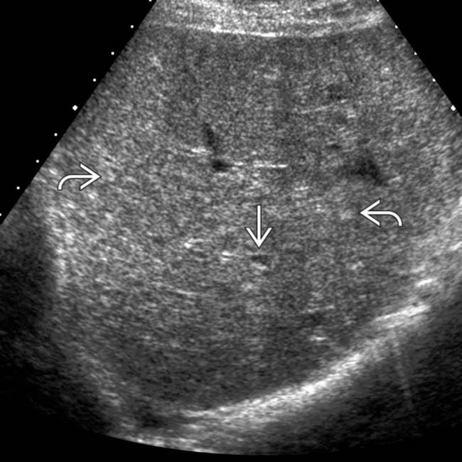
 throughout the liver and 1 of ∼ 10 cyst-like lesions
  , though even these have small foci of echogenicity within the wall. MR on this patient showed many more cystic-appearing biliary hamartomas.