Biological and pathological introduction

Chapter 16 Biological and pathological introduction


Radiotherapy is used almost exclusively for the treatment of cancer and related conditions. It is thus very important to have a reasonable understanding of this disease. This chapter contains an outline of the basic characteristics of cancerous cells, and the conditions that can cause cancer. There is consideration of the natural history of untreated cancer, and of the ways that different types of cancer are named and classified.


Growth: proliferation, differentiation and apoptosis


Growth is the process of increase in size and maturity of tissues from fertilization through to the adult. When normally controlled, the different parts of the body take on their correct size and specialist functions, and relationship to one another. Furthermore, throughout life these attributes continue despite the need for replacement and repair. This is all a reflection of accurate control of the timing and extent of cellular proliferation, cell-to-cell orientation and organization, and differentiation. Differentiation is the process of a cell taking on a specialized function; this is usually associated with a change in its microscopic appearance. It is also usually a one-way commitment, with relative or complete loss of the ability to continue proliferating. Also important is the ability to delete cells which are no longer needed, by a process called apoptosis or programmed cell death. This is used in the development of the fetus, and in maintaining or adjusting the size of a structure in the adult. Apoptosis is also used to delete defective cells.


Neoplasia


A neoplasm can be defined as a lesion resulting from the autonomous or relatively autonomous abnormal growth of cells which persists after the initiating stimulus has been removed; i.e. cell growth has escaped from normal regulatory mechanisms. The abnormality affects all aspects of cell growth and apoptosis to varying degrees. Proliferation continues unabated, irrespective of the requirements of the organ in which the neoplasm is situated. This, combined with loss of control of the normal relationships between cells, often results in the new tumour cells replacing and insinuating themselves between the adjacent normal tissues, a process called invasion. Loss of differentiation accompanies, and often correlates with, failure of proliferation control and invasiveness. Failure of apoptosis may also be a major contribution to the survival of abnormal cells.


Benign and malignant neoplasms


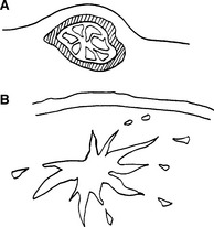
Neoplasia is not a single disease, but rather a common pathological process with a multitude of different varieties and clinical outcomes. One fundamental division is between benign and malignant tumours. Benign tumours will remain localized, with generally relatively little effect on the patient. In contrast, other tumours are locally destructive, may spread to involve other parts of the body, and ultimately result in the death of the patient. Figure 16.1 and Table 16.1 show some of the differences between benign and malignant neoplasms. Further aspects of the classification of tumours are discussed later in this chapter.


Table 16.1 Characteristics of neoplasms


Feature Benign Malignant
Growth rate Slow Variable, may be rapid
Margin Encapsulated Invasive
Local effect Little Destructive
Differentiation Good Variable, may be poor
Metastases No Frequently
Usual outcome Good Fatal

The term cancer (Latin for ‘crab’) is very ancient, and there are several explanations for its usage. Some say it reflects the tenacious grip the disease has on its victim, some that it describes the radiating prominent veins that may surround an advanced superficial tumour. Others contend that it describes the irregular infiltrative profile of some tumours, e.g. of breast. Suffice it to say that cancer is a common colloquial term that is generally applied to any malignant neoplasm.


Though the behaviour of tumours, particularly malignant ones, may seem very odd, it can generally be explained by the excessive or inappropriate expression of genes that are present in all cells. The tumour continues to be dependent upon an adequate blood supply (though many acquire the capacity to induce new vessels). Also, many of those which arise from hormone-dependent tissues (e.g. breast, prostate) continue to show a degree of dependence on those hormones. This can be exploited to therapeutic advantage by giving antihormone treatment.


Carcinogenesis


The causes of cancer are numerous, and mechanisms of its production are complex, but some of the principles will be presented. There are two avenues of thought to follow: one at the molecular and genetic levels, the other concerned with causative associations. It is not always easy to relate these to each other. Underlying the mechanistic approach is the assumption that cell behavior is controlled by the genes expressed (which ones, and how strongly), bearing in mind that these can be influenced by chemical messages relayed from outside the cell. Every cell contains the entire genetic code, but only expresses those genes appropriate to its own situation. In cancer, this has gone wrong, particularly with respect to proliferation, differentiation or apoptosis. We can generalize to say that multiple abnormalities need to have occurred between normality and cancer, and that this reflects a multistep process. Only those cells capable of division are at risk of transforming into a neoplasm. This excludes terminally differentiated cells such as circulating red cells, the uppermost keratinized cells of the skin, and adult voluntary muscle and nerve cells.


Oncogenes and tumour suppressor genes


The function of a cell is critically dependent on the expression of its genes. In cancer, it has been observed that there may be both inappropriate levels of expression of otherwise normal genes, and abnormalities of genes. Though copying of the genetic code from one cell generation to another is very accurate, it is not perfect. An abnormal copy or mutation may give rise to a protein with excessive function or reduced function, or may fail to produce a protein at all, resulting in no function. Alternatively, it may be the control of the gene which is altered, so that there is increased or decreased expression resulting in excess or deficient protein product and hence function. An oncogene is an altered gene which contributes to cancer development when its expression is increased. The normally functioning counterparts of these genes are often concerned with control of cell proliferation.


In contrast, tumour suppressor genes are those which, when totally absent or non-functioning in a cell, permit the emergence of neoplasia, i.e. their presence prevents neoplasia. Given that all normal cells will have two copies of each gene (one on each chromosome pair), the development of neoplasia by this means requires loss or mutation of both copies.


It must be stressed that clinical cancer does not reflect a solitary abnormality of one of these genes, but rather the final result of a combination of several errors of function. The gradual accumulation of multiple genetic defects is typified by the progression of a benign polyp in the colon to a cancer over about 10 years. How is it that a cell can acquire so many abnormal genes? The explanation in several situations is that there is failure of the screening of the cell’s genetic code for abnormalities, or defective repair of incidental genetic damage. The protein p53 is important in assuring the integrity of the genome, and it is frequently defective in cancer cells. Similarly, the failure of DNA repair can result in much more rapid accumulation of mutations – the mutator phenotype.


Defective apoptosis has several consequences with regard to cancer. Inappropriately increased expression of BCL-2, which inhibits apoptosis, is a major reason for cell accumulation in some tumours, for example follicular lymphoma. Failure of apoptosis may contribute to survival of defective cells and also cell survival in abnormal environments such as occurs during invasion and metastasis. Finally, many cancer treatments rely on induction of apoptosis to kill tumour cells, so these will be less effective if apoptosis is defective.


Physical agents


Ionizing radiation


There is no doubt that ionizing radiation can cause cancer. Direct damage to DNA (i.e. the chemical basis of genetic information), and damage mediated via ionization of water can result in mutation of genes. The damage is randomly scattered throughout the genetic code, but can include sites critical to the development of cancer by the usual sequence of initiation, promotion and so on. Traditionally, it has been thought that radiation causes a somatic mutation in a normal cell. The earliest event is probably genomic instability. Subsequently, there is a multistep sequence of genetic events. This typically results in an interval of many years between exposure and clinical cancer. The source of radiation does not matter from the point of view of causing cancer, though it will affect the sites at risk.


Ionizing radiation can lead to loss of tumour suppressor genes and activation of proto-oncogenes. Oncogenes may also be activated as result of point mutations. Gene amplification can lead to activation and overexpression of a proto-oncogene. It had previously been thought that mutagenesis only occurred in normal cells traversed by radiation particles. However, normal cells can undergo change without such damage by virtue of what is termed the ‘bystander effect’. The mechanism of this bystander effect is not clear but it could be due to secretion of factors (as yet unidentified) from irradiated cells that influence the survival of adjacent non-irradiated cells.

Stay updated, free articles. Join our Telegram channel

Mar 7, 2016 | Posted by in GENERAL RADIOLOGY | Comments Off on Biological and pathological introduction

Full access? Get Clinical Tree

Get Clinical Tree app for offline access