Gastrointestinal cancer

Chapter 24 Gastrointestinal cancer


Chapter contents


Cancer of the oesophagus


Stomach


Hepatocellular carcinoma


Pancreas


Colon and rectum


Anal cancer


Further reading


Cancer of the oesophagus


Anatomy (Figure 24.1)
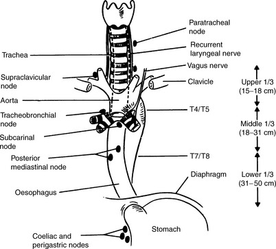

The oesophagus is a flattened muscular tube of 18–26   cm in length with diameters varying from 1.6 to 2.5   cm. The oesophagus starts at the pharyngoesophageal junction at the level of C5–6 vertebral interspace and ends at the esophagogastric junction at the level of T1 vertebra. It is traditionally divided into three segments, cervical, thoracic and abdominal. The cervical segment is about 4–5    cm long and extends from the pharyngoesophageal junction to the thoracic inlet. The thoracic oesophagus extends from the thoracic inlet to the diaphragmatic opening. The abdominal segment is short varying from 1 to 2.5   cm, extending from the hiatus to the esophagogastric junction. The esophageal hiatus in the diaphragm is at the level of T10 vertebra. The oesophagus moves a few millimeters during quiet respiration and by as much as 3–4   cm during swallowing.


The lymphatic drainage is of importance to the surgeon and the oncologist. A dense lymphatic network is found in the submucosa, which travels a variable distance longitudinally before penetrating the muscle layer and draining into the periesophageal lymph nodes. This may explain the extensive intramural spread of the cancer beneath an intact mucosa and tumour-free margins may not necessarily mean complete tumour resection. The lymphatic drainage of the proximal oesophagus is towards the deep cervical chain and thoracic duct. The distal oesophagus drains into the lower mediastinal, left gastric and coeliac axis nodes.


The esophageal wall is composed of four layers: the innermost non-keratinized squamous epithelium – the mucosa, submucosal connective tissue, muscularis propria with inner circular and outer longitudinal layers and, finally, the adventitial layer. The myenteric or Auerbach plexus of nerves responsible for coordinating swallowing is located between the inner circular and outer longitudinal layers. The oesophagus has no mesentery or serosal lining which may explain the relatively easy spread of esophageal cancer into the surrounding tissues.


Diagnostic evaluation (Tables 24.124.3)


Any patient who presents with recent onset dysphagia and weight loss should be investigated for a possible esophageal cancer. Many major centres have a fast track dysphagia endoscopy service available to minimize the delay in the diagnosis of the cancer.


Table 24.1 Staging esophageal carcinoma


TNM staging system applies only for esophageal carcinomas
T: Primary tumour
Tx Primary tumour cannot be assessed
T0 No evidence of primary tumour
Tis Carcinoma in situ
T1 Tumour invades the lamina propria or submucosa
T2 Tumour invades the muscularis propria
T3 Tumour invades the adventitia
T4 Tumour invades the adjacent structures
N: Regional nodes (see below)
NX Regional lymph nodes cannot be assessed
N0 No regional lymph node metastases
N1 Regional lymph node metastases
M: Distant metastases
MX Distant metastases cannot be assessed
M0 No distant metastases
M1 Distant metastases
For tumours of the upper thoracic oesophagus
M1a Metastases in cervical lymph nodes
M1b Other distant metastases
For tumours of the mid-thoracic oesophagus
M1a Not applicable
M1b Non-regional lymph nodes or other distant metastases
For tumours of the lower thoracic oesophagus
M1a Metastases in coeliac axis lymph nodes
M1b Other distant metastases
The regional lymph nodes depend on the anatomical subsite of the oesophagus (cervical or intrathoracic)
Cervical oesophagus
Scalene, internal jugular, upper and lower cervical, periesophageal and supraclavicular nodes
Intrathoracic oesophagus
Upper periesophageal (above the azygos vein), subcarinal, lower periesophageal (below the azygos vein), mediastinal, perigastric except coeliac axis nodes
pTNM Pathological classification


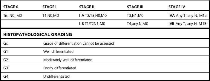
Table 24.3 Diagnostic evaluation of patients with esophageal cancer


Full blood count & renal & liver chemistry


CT scan of the chest & abdomen


Endoscopic ultrasound (EUS ± FNA)


Bronchoscopy (in selected cases)


FDG PET-CT


Isotope bone scan (if indicated)


In the majority of cases, upper GI endoscopy visualizes the tumour and its distance from the incisor teeth. The endoscopist should also define the lower extent of the tumour (if the lumen is traversable) and evaluate the esophagogastric junction. Multiple biopsies should be taken to maximize the yield. Any associated Barrett’s segment, its length and relation to the invasive cancer should be noted.


In patients who are fit for curative treatment, the first step is a multislice computed tomography (CT) scan of the chest and abdomen to exclude obvious metastatic disease. CT has poor sensitivity in predicting the T or N stage. The sensitivity and specificity of CT for T4 disease are 25% and 94% respectively and for coeliac node involvement are 30% and 90% respectively.


In patients with localized disease, further evaluation of the T and N stage is best done by endoscopic ultrasound (EUS) by an experienced endoscopist. EUS has a high sensitivity and specificity in evaluating the T and N stage in esophageal cancer and has improved the preoperative staging of esophageal cancer. The overall accuracy of EUS is 85% for T staging and 75% for N staging. EUS fine needle aspiration (FNA) enhances the accuracy of nodal staging and, in particular, is useful in confirming coeliac axis nodal status on cytology. EUS is less reliable in assessing T and N stage following preoperative chemoradiation because it may not be able to differentiate between residual tumour and post-radiation inflammation/fibrosis.


In patients with hoarseness of voices or hemoptysis/unexplained cough, bronchoscopy is recommended to rule out recurrent laryngeal nerve involvement or tracheobronchial fistula.


The role of 2-fluorodeoxy-D-glucose positron emission tomography (FDG PET-CT) in the initial staging of potentially operable esophageal cancer has been the subject of numerous studies. In a recent systematic review, the pooled sensitivity and specificity of FDG PET for distant nodal and hematogenous metastases was 67% and 97%, respectively. The change in patient management as a result of FDG PET ranged from 3 to 20%. Preliminary data suggest that early FDG PET following neoadjuvant chemotherapy may predict pathologic response and overall survival and thus may define a subset of patients who are more likely to benefit from surgery.


Radiation therapy techniques


External beam radiation


Radiation planning using oral contrast (barium or gastrograffin) underestimates the tumour length. Three-dimensional conformal radiotherapy with CT planning is recommended. Joint planning with the endoscopist/radiologist to delineate the primary tumour based on the endoscopic ultrasound information will help to minimize the risk of a geographic miss. EUS can detect submucosal spread and gives a more accurate extent of the disease. Conventional margins above and below the tumour used to be 5   cm but, with modern imaging, this margin can be reduced to 3   cm and a 1.5–2   cm margin around the gross tumour volume (GTV) is adequate. A 3D conformal plan is generated with dose volume histograms defining the doses for the planned target volume, regions of interest including the spinal cord and lungs. A single phase three-field (anterior and two obliques) is the standard field arrangement to deliver a dose of 45–50   Gy in 1.8–2   Gy fractions. Chemoradiation patients will have concomitant intravenous 5-fluorouracil (5FU) or oral capecitabine and cisplatin during their radiation treatment. A widely used regimen is cisplatin 75   mg/m2 day 1 plus 5-fluorouracil 1000   mg/m2 infused over 24 hours on days 1–4 repeated every 3 weeks. Two to four cycles are given during radiotherapy. Weekly blood count and renal function monitoring is mandatory. Continuing dietary assessment prior to, during and after radiation, with nutritional support if necessary, is strongly recommended. Parallel-opposed fields (anterior/posterior) can be used for palliative radiotherapy.


Stomach


Staging (Tables 24.4 and 24.5)


The initial investigations often include a barium meal/swallow and endoscopy. Gastroscopy and biopsy will confirm the diagnosis in the majority of patients. Multiple biopsies increase the sensitivity and yield. The TNM staging system is used for staging gastric cancer. Endoscopic ultrasound is of value in proximal cardia and junctional tumours. Cross-sectional imaging with a spiral CT is the first step to exclude overt metastatic disease. CT is not a sensitive tool for detecting peritoneal spread, therefore, laparoscopy is mandatory prior to surgery to detect any occult peritoneal spread. Laparoscopy may also help in identifying tumour in the lesser sac, on the surface of the liver and nodal disease. FDG-PET imaging may identify metastases not clearly seen on CT but 25–30% of primary gastric cancers are not FDG avid.


Table 24.4 Stomach cancer TNM clinical classification


Only gold members can continue reading. Log In or Register to continue

Stay updated, free articles. Join our Telegram channel

Mar 7, 2016 | Posted by in GENERAL RADIOLOGY | Comments Off on Gastrointestinal cancer

Full access? Get Clinical Tree

Get Clinical Tree app for offline access
T: Primary tumour
TX Primary tumour cannot be assessed
T0 No evidence of primary tumour
Tis Carcinoma in situ: intraepithelial tumour without invasion of the lamina propria
T1 Tumour invades lamina propria or submucosa
T2 Tumour invades muscularis propria or subserosa
T3 Tumour penetrates serosa (visceral peritoneum) without invasion of adjacent structures
T4 Tumour invades adjacent structures
Notes:

.
N: Regional lymph nodes
NX Regional lymph nodes cannot be assessed
N0 No regional lymph node metastasis
N1 Metastasis in 1 to 6 regional lymph nodes
N2