Thyroid cancer

Chapter 23 Thyroid cancer


Anatomy


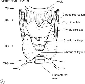
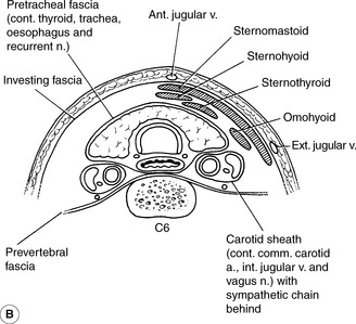
The thyroid gland is situated in the anterior part of the neck just above the clavicle and sternum (Figure 23.1). The gland consists of a right and left lobe joined by an isthmus which crosses the trachea at the second and third cartilaginous rings. The average weight of the thyroid gland is 20   g. The parathyroid glands lie on the posterior surface of both thyroid lobes and the recurrent laryngeal nerves are in a cleft between the trachea and the oesophagus. Lymphatic drainage from the thyroid can be to nodes superior to the thyroid gland, lateral to the gland and to the paratracheal region.


Etiological factors


Thyroid cancer shows two age-related peaks of incidence, one before the age of 40 and one after 60. The condition is approximately three times as common in women than men. Exposure to ionizing radiation in childhood, either therapeutic, such as radiotherapy for Hodgkin’s disease, or associated with environmental exposure, increases the risk of developing thyroid cancer. There is, however, no proven relationship between the development of thyroid cancer and administration of iodine-131 for the treatment of hyperthyroidism.


Other medical conditions which affect the thyroid have also been linked with an increased risk in developing thyroid cancer, namely, endemic goiter, which occurs in areas where there is low natural iodine, Hashimoto’s thyroiditis, a past history of thyroid adenoma or a family history of thyroid adenoma or cancer. Some genetic conditions which increase the risk of thyroid cancer include Gardener syndrome, Cowden’s syndrome, familial adenomatous polyposis and the multiple endocrine neoplasia syndromes.


Thyroid cancer initiation and progression is supposed to be through accumulation of genetic and epigenetic events. It point mutations of BRAF (Predominantly Papillary) and RAS (N,H,K-Ras Predominantly Follicular) genes and Chromosomal rearrangements-RET/PTC-(Papillary particularly radiation induced), PAX8/PPARγ (Follicular); abnormal miRNAs’ (microRNA) with gene methylation problems also play its part. All these are now being detected on Fine needle aspiration cytology. This will avoid diagnostic lobectomies for indeterminate and benign nodules in the future protecting patients from unnecessary surgery as well as allowing one-stage total thyroidectomy for cancer patients (instead of lobectomy followed by total thyroidectomy in Thy3 cases) and saving costs. P53 mutations are thought to be a late event in progression from differentiated to Anaplstic thyroid cancer in a high proportion of cases.


Presentation, diagnosis and patient pathway


The most common presenting feature is a painless, lower neck lump in the thyroid area which may or may not have been noted to have increased in size. In more advanced cases, patients may complain of a lump on the side of the neck due to spread to lymph nodes, hoarseness or change in voice suggesting recurrent laryngeal nerve involvement, stridor or, rarely, dysphagia. A rapid enlargement of the mass or a rapid development of symptoms, suggesting compression of other structures, may be due to a more rapidly growing anaplasic carcinoma or Lymphoma.


In the first instance, the patient should be referred to an ENT or general surgeon with a particular interest and experience in managing thyroid cancer. A history of the development of the mass, any relevant personal past history or family history of thyroid problems is important. Examination is undertaken to define whether the mass is solitary or multiple, whether it is tender, which might suggest a benign cause, and whether the mass is fixed to the skin or to underlying structures in the neck. Any enlarged lymph nodes within the neck are noted. Development of stridor is a medical emergency, demanding urgent hospital admission.


Ultrasound and Fine Needle Aspiration Cytology (FNAC) are essential for neck masses suspected of being a thyroid cancer. Ultrasound guidance may help in obtaining the aspiration cytology and can help to distinguish solid from cystic masses in the neck. It can differentiate between solitary and multiple nodules, helps to detect involved lymphnodes (if needed by guided FNAC) and can provide sonographic features suggesting probable malignancy in some cases.


Follicular cancers cannot be differentiated from adenomas on FNAC alone as evidence of invasion is required and lobectomy is usually necessary. If FNA cytology is not helpful, it should be repeated and, if this is still inconclusive, thyroid lobectomy should be performed to obtain enough tissue for diagnosis. There are no indications for an incisional biopsy of the thyroid, which might compromise future therapeutic interventions.


Radioactive iodine scans can rarely help differentiate non-functioning ‘cold’ nodules within the thyroid, which may represent a malignancy, from ‘hot’ nodules which rarely contain a cancer, but usually these scans are not helpful with diagnosis, and are much more useful in the postoperative period for diagnosing residual disease or early recurrence.


Computed tomography (CT) and magnetic resonance imaging (MRI) scans are not routinely required but can help show the local extent of spread, detailing involvement of lymph nodes and other neck structures.


Thyroid function tests, which are usually normal in patients with thyroid cancer, calcium, phosphate and thyroglobulin can be sampled for baseline measurement, but thyroglobulin level is not useful in the preoperative phase. If there is any suspicion that the thyroid tumour may be a medullary cancer, then the calcitonin level is checked.


Staging is performed using the TNM system which is summarized in Table 23.1. All anaplastic carcinomas are considered T4 tumours.


Table 23.1 TNM staging system


T1 Tumour <=2   cm, intrathyroidal
T1a <=1   cm
T1b 1-2   cm
T2 Tumour >2-4   cm, intrathyroidal
T3 Tumour >4   cm on with minimal extra-thyroid extension
T4 Tumour extending beyond the thyroid capsule
T4a Subcutaneous tissue, larynx, trachea, R L Nerve. Oesophagus
T4b Prevertebral fascia, mediatinal vessels, carotid artery
N0 No regional lymph node involvement
N1 Regional lymph node involvement
N1a Level VI
N1b Other regional
M0 No distant metastasis
M1 Distant metastasis present

One major disadvantage of the TNM system is that the presence of positive nodes does not necessarily give a worse prognosis, consequently making this staging system less useful in the practical setting than at other cancer sites.


In addition to the TNM system, there are other risk categorisation systems. ‘AMES’, based on age, distant metastasis, tumour extent and tumour size.


This system defines high-risk patients as:


Low-risk patients are described as:


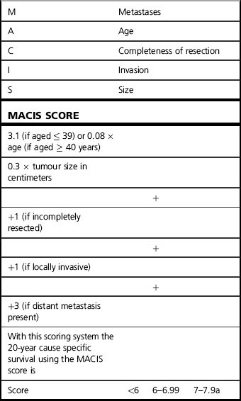
Another system which gives prognostic information is the MACIS scoring scheme developed at the Mayo Clinic (Table 23.2).


Differentiated thyroid cancer


These tumours are divided into papillary and follicular thyroid cancers and are distinguished by their behavior, as they are sensitive to thyroid stimulating hormone (TSH), take up iodine and can produce thyroglobulin. All three of these factors are used in their management. It should be remembered that, in general, differentiated thyroid cancer carries a very good prognosis, with 20-year survival of over 85% in those patients who do not develop distant metastasis.


Papillary thyroid cancer is the most common type of thyroid cancer, accounting for almost 80% of all cases and its incidence is increasing world wide. These tumours are also derived from the follicular cells of the thyroid, and may show psammoma bodies, small flecks of calcification, on microscopy. The distinctive feature of these tumours is that they have characteristic nuclei (Orphan-Annie appearance) and they seem to grow in branching stocks (the so-called papillae). These tumours are often multifocal and they can show more local extension and cervical node involvement and less tendency to haematogenous spread when compared to follicular cancers. Papillary carcinoma is three times more common in women than men with a peak incidence in the third to fourth decade of life.


Follicular thyroid cancer makes up about 10% of thyroid malignancy. It generally affects a slightly older age group than papillary cancer and tends to involve the neck nodes less. Follicular carcinomas are often solitary within the thyroid gland, have a marked tendency to invade the vascular channels and to spread via the bloodstream to distant sites.


Surgery


Surgery is required for all thyroid cancer, if operable (except Lymphoma). If differentiated thyroid carcinoma >1   cm in size is confirmed, total thyroidectomy or near total thyroidectomy, with preservation of the recurrent laryngeal nerves and at least some of the parathyroid glands, should be carried out depending on diagnostic findings. Total lobectomy alone is indicated rarely in <1   cm or very low risk tumours. Surgical should be individualised and based on clinical, sonographic, staging and cytology findings.


Nomenclature currently used for diagnostic categorization is the Thy 1, Thy 2 etc. system, and can be found in the ‘Guidelines for the management of Thyroid Cancer in Adults’ published by the British Thyroid Association and the Royal College of Physicians of London. This system can be summarized as shown in Table 23.3. Further refinements of this system into Thy3a (atypia) and Thy3f (Follicular lesion)have been proposed recently.


Table 23.3 British Thyroid Association coding system


Only gold members can continue reading. Log In or Register to continue

Stay updated, free articles. Join our Telegram channel

Mar 7, 2016 | Posted by in GENERAL RADIOLOGY | Comments Off on Thyroid cancer

Full access? Get Clinical Tree

Get Clinical Tree app for offline access
Thy1 Non-diagnostic; FNAC needs repeating
Thy2 Non-neoplastic; two diagnostic tests with benign results with 3–6 months apart are needed to exclude malignancy
Thy3 All follicular lesions; lobectomy required with completion thyroidectomy if malignant histology