Case presentation
An 11-year-old male presents with left wrist pain and swelling 3 days after he fell while playing soccer. Apparently, in the course of the game, he fell on his outstretched left arm. After the injury, he continued to play but later in the day he noted pain and swelling to the left wrist. He denies neck, shoulder, elbow, upper arm, hand, or digit pain. He denies back pain.
Physical examination reveals a well-appearing patient, in no distress, with age-appropriate vital signs. He has obvious swelling to the left wrist and there is pain with palpation of the distal radius and ulna; this pain is reproduced with movement of the wrist. He has no shoulder, upper arm, elbow, or hand tenderness or swelling and there is full range of motion at these areas. His radial pulse is strong with good capillary refill. He is grossly neurovascularly intact.
Imaging considerations
Plain radiography
This is the initial imaging testing of choice for suspected wrist fractures. Exposure to ionizing radiation is low and the modality is readily and widely available. Two views (anteroposterior and lateral) should be obtained, although an oblique view may be helpful if a fracture is suspected but cannot be completely visualized on two-view imaging.
Ultrasound
This modality is advantageous given the lack of radiation and rapidity. Ultrasound has been demonstrated to be effective in identifying wrist fractures, including when performed by physicians with minimal ultrasound experience.
Computed tomography
This modality is not initially employed but may be utilized to better visualize complex fractures. Consultation with a pediatric orthopedic specialist is appropriate prior to obtaining computed tomography imaging.
Magnetic resonance imaging (MRI)
This modality is not a first-line imaging test. If there is a complex fracture, concern for neurovascular compromise, concern for an infectious process, or concern for an oncologic process, use of MRI may be appropriate. Consultation with a pediatric orthopedic specialist is appropriate prior to obtaining MRI.
Imaging findings
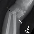
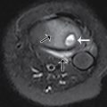
The patient had plain radiography of his wrist, including anteroposterior, lateral, and oblique views. These images demonstrate a subtle Salter-Harris type II fracture of the distal left radius with minimal posterior displacement of the distal fragment; there is also a Salter-Harris type I fracture of the distal left ulna that is anatomically aligned. Soft tissue swelling surrounds the fracture sites ( Figs. 59.1 and 59.2 ).


Case conclusion
The patient was placed in a sugar tong splint and arrangements were made to follow-up with Pediatric Orthopedics. He kept his appointment 1 week later and at that time was placed in a short arm cast. Four weeks later, the cast was removed and a Velcro wrist splint was applied, which was worn for several weeks more. He recovered uneventfully. Interestingly, he was seen approximately 1 year later for a second left wrist injury he sustained while falling during a football game. Imaging at this visit demonstrated a new, acute Salter-Harris type II fracture of the left distal radius with posterior displacement of the distal fragment ( Fig. 59.3 ). This time, the fracture was reduced in the emergency department by Pediatric Orthopedics under sedation, a cast was placed, and the patient was discharged with close observation and follow-up.


